
A 57-year-old Hispanic female who recently moved to Miami presented complaining of poor vision in her left eye for several years. Her last eye exam was in Cuba about two years earlier. At that time, she was told that a suspicious lesion in her left eye required both laser and a freezing treatment. The patient was concerned about the status of her left eye. She said she has not had any problems with her right eye.
Her medical history is significant for hypertension and type I diabetes, for which she takes lisinopril, glyburide and Xanax (aprazolam, Pfizer).
On examination, her best-corrected visual acuity measured 20/20 in the right eye and light perception in the left. Her motility testing was normal.
Confrontation visual fields were full to careful finger counting in the right eye but were grossly constricted to only light perception in the left. There was a strong afferent pupillary defect in the left eye. Her anterior segment exam was significant for 1+ nuclear sclerotic cataract in both eyes.
Dilated fundus exam was normal in the right eye. The findings in the left eye are shown (figure 1). Standardized echography performed showed a lesion that measured 12.5mm x 9.5mm x 2.4mm in elevation (figure 2).
 |
| 1. This is a retinal montage of the fundus in the patients left eye. Note the lesion in the posterior pole that is temporal to the macula. Also, note the secondary pigmentary change that overlies and surrounds the lesion. |
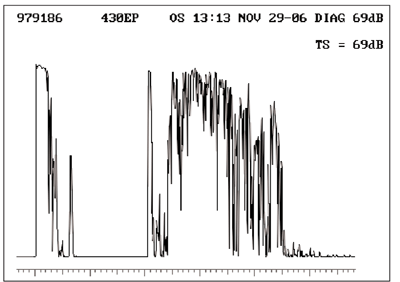 |
| 2. The diagnostic A-scan of our patient shows an initial highly reflective spike that represents the retina, followed by a group of lower reflective spikes that likely represent subretinal opacities. |
Take the Retina Quiz
1. Based on the A-scan, what is the reflectivity of the lesion?
a. Low.
b. Medium.
c. High.
d. Unable to determine due to the scarring.
2. What is the correct diagnosis?
a. Choroidal nevus.
b. Uveal melanoma.
c. Choroidal hemangioma.
d. Hemorrhagic detachment of the retinal pigment epithelium (RPE) secondary to choroidal neovascularization.
3. What other secondary changes does the fundus photograph reveal?
a. Metastasis to the retina.
b. Exudative retinal detachment.
c. Macular disciform scar.
d. Bad case of eye rot.
4. How should this patient be managed?
a. Observation.
b. Enucleation.
c. Plaque radiotherapy.
d. Photodynamic therapy (PDT).
5. What is the visual prognosis of this eye?
a. Very poor.
b. Moderate.
c. Excellent.
d. Uncertain.
For answers, see below.
Discussion
Our patient has a nonpigmented 2.4mm elevated lesion temporal to the macula. Secondary RPE hyperplasia overlies and surrounds the lesion. There is also an overlying exudative retinal detachment that extends close to the optic nerve. The fundus photograph shows the demarcation line of the retinal detachment adjacent to the optic nerve.
Clearly, the lesion looks suspicious and could be a malignancy, such as a uveal (choroidal) melanoma. Areas of atrophy around this patients lesion and the RPE hyperplasia are not typical of a melanoma; they are more a sign of chronicity. Even so, we must rule out a melanoma, which is best done with ultrasound.
The diagnostic A-scan of our patient shows an initial highly reflective spike that represents the retina, followed by a group of lower reflective spikes that likely represent subretinal opacities. The next group of spikes represents the tumor; the first spike is highly reflective followed by a tighter group of spikes that have medium to highly reflectivity. As the A-scan moves posteriorly, the remaining spikes are highly reflective; these represent orbital fat.
The lesions high internal reflectivity is the significant finding. This is typical for a choroidal hemangioma with a secondary exudative retinal detachment.
Choroidal hemangioma is a benign and relatively rare vascular tumor. It can present as a discrete, well-circumscribed lesion or as a diffuse tumor. The diffuse form can be very large and is most often associated with Sturge-Weber syndrome. Clearly, our patient has the more circumscribed form.
We can often diagnose choroidal hemangioma based on the clinical appearance. The color of the lesion is the diagnostic feature. Choroidal hemangiomas usually appear pink or orange-red, but they can also appear gray, yellow or even flesh colored.
These tumors are rarely as elevated and prominent as our patients tumor. More commonly, they can be flat or minimally elevated.
The less distinct tumors can often be missed on clinical exam, as they can blend almost imperceptibly with the surrounding choroidal tissue. Because of this, they may be more visible with binocular indirect ophthalmoscopy (BIO) instead of slit lamp biomicroscopy. The higher magnification of the slit lamp doesnt easily allow for the detection of the sometimes subtle color changes between the retina and the tumor.
Choroidal hemangiomas are commonly located in the posterior pole, most often within the paramacular area. Most patients are asymptomatic, and these tumors may be discovered as an incidental finding during an exam. Symptoms may develop if the lesion is located directly under the macula, or if the patient develops a neurosensory detachment that involves the macular area; this is what occurred in our patient.
Standardized ultrasound can be very helpful in establishing or confirming the diagnosis of choroidal hemangioma vs. other similarly appearing lesions, such as a uveal (choroidal) melanoma. On A-scan, there will be a high initial spike that corresponds to the anterior tumor surface followed by several medium to highly reflective spikes within the tumor, as seen in our patient. Choroidal melanoma, however, typically has low to medium reflectivity.
In the absence of subretinal fluid involving the macula, most of these tumors can be managed with observation alone. The presence of fluid in the macula warrants surgical intervention.
Over the years, several different treatments have been advocated. Laser photocoagulation has been the mainstay. The idea is to lightly treat the surface of the lesion with the hope of collapsing the cystic retina onto the surface of the tumor, ultimately resolving the subretinal fluid.
More recently, photodynamic therapy (PDT) has emerged as the treatment of choice for choroidal hemangioima. These tumors seem to respond very well to the effects of the nonthermal laser along with Visudyne (verteporfin, Novartis Ophthalmics).1 The treatment effect can be enhanced when combined with intravitreal Kenalog (triamcinolone, Bristol-Meyers Squibb) injection (IVK).
Our patient underwent PDT with IVK to try to shrink the tumor and resolve the exudative retinal detachment. Because of the chronicity of the lesion, the secondary retinal changes and the profound vision loss, the visual potential of this eye is very poor. Still, we hope that resolution of the retinal detachment will allow for some functional vision in this eye.
Meanwhile, we recommended precautions for her other eye, including spectacles with polycarbonate lenses. We continue to follow the patient.
1. Sanborn GE. Circumscribed choroidal hemangioma. In: Ryan SJ (editor-in-chief). Retina, Vol. IIMedical Retina. Tumors of the Retina, Choroids and Vitreous. 4th ed. St. Louis: Mosby;2006:829-42.
Retina Quiz Answers: 1) c; 2) c; 3) b; 4) d; 5) a.
