A 39-year-old Haitian female presented to the emergency room with headaches and decreased vision in both eyes. The patient reported that she had experienced a gradual, painless loss of vision during the last month; however, the symptoms became especially severe during the past four days.
Her medical history was significant for HIV, HIV encephalopathy, Mycobacterium avium complex (MAC) and hypertension. A caregiver who accompanied her to the office said that the patient recently was hospitalized for altered mental status and arm weakness. At that time, her CD4 count was 11. Her current medications included ethambutol, azithromycin and lisinopril.
On examination, we noted resolving vesicular lesions on the left side of her face, which did not appear to involve her eyes. Her entering visual acuity was light perception only O.U. Her pupils measured 8mm in each eye and were nonreactive. Motility testing was full. The anterior segment examination showed trace cell in the anterior chamber O.U. There were also trace to 1+ anterior vitreous cells in both eyes. There were no iris nodules. Her intraocular pressure measured 16mm Hg O.U.
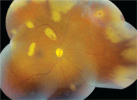
1. Here is a peripheral montage of the patient’s right eye. Note the peripheral and posterior pole lesions.
Dilated fundus exam showed pale nerves with moderate-sized cups O.U. She had obvious circumferential peripheral white lesions in her right eye (figure 1). We also documented several focal white lesions scattered throughout the right
posterior pole. In the left eye, we noted similar peripheral lesions as well as two smaller focal lesions that were located outside the posterior pole (figure 2).
Take the Retina Quiz
1. How would you categorize the retinal lesions seen in our patient?
a. Infectious.
b. Inflammatory.
c. Metastatic.
d. Immune mediated.
2. What is the likely diagnosis?
a. Cytomegalovirus (CMV)
retinitis.
b. Necrotizing herpetic retinitis (NHR).
c. Syphilis.
d. Active toxoplasmosis.
3. Which test(s) would be most appropriate for this patient?
a. Serologic studies, including FTA-ABS, rapid plasma reagin (RPR) and toxoplasmosis IgG/IgM.
b. Lumbar puncture (LP).
c. Anterior chamber tap with polymerase chain reaction (PCR).
d. All of above.
4. What is the appropriate treatment for this patient?
a. Anti-VEGF therapy.
b. Antiviral therapy.
c. Intramuscular penicillin.
d. Oral Bactrim (sulfamethoxazole/trimethoprim, URL Pharma).
5. What is the prognosis for visual recovery?
a. Very poor.
b. Fair.
c. Very good.
d. Unknown.
For answers, see below.
Discussion
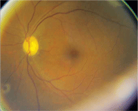
2. The posterior pole of the patient’s left eye. What do you see?
With a CD4 of 11, it is evident that our patient is severely immunocompromised. Even worse, she has profound vision loss secondary to optic atrophy as well as an unusual retinal condition. After careful examination, the lesions appeared to be at the level of the retina and retinal pigment epithelium (RPE). With the presence of vitreous cells, we believed that this presentation was likely infectious in origin. Given their peripheral circumferential configuration, viral necrotizing retinitis––either acute retinal necrosis (ARN) or progressive outer retinal necrosis (PORN)—ranked very high on our list of differential diagnoses. Additionally, the resolving vesicular changes on the left side of her face suggested the possibility of a herpes zoster infection.
We admitted the patient to a local hospital where she underwent a series of tests. She had serologic studies, including CBC, toxoplasma IgG/IgM antibodies, angiotension converting enzyme (ACE), lysozyme, RPR, FTA-ABS and cryptococcal studies. She also had a PPD and chest X-ray, CT and MRI, as well as a LP and anterior chamber tap using PCR analysis of aqueous humor samples to examine for herpes zoster virus (HZV), type 1 and type 2 herpes simplex virus (HSV), CMV, Epstein-Barr virus (EBV) and Toxoplasma antibodies. While waiting for the results, we began the patient on induction doses of intravenous foscarnet and ganciclovir. When the results came back, the anterior chamber PCR tap was positive for varicella zoster virus (VZV).
Necrotizing herpetic retinitis refers to a spectrum of diseases induced by herpes viruses, such as HSV-1 and HSV-2, VZV, CMV or EBV.1,2 Though all extremely rare, the two primary retinal conditions that are commonly seen within the herpes family are ARN and PORN. Both conditions tend to progress rapidly (days to weeks), and usually begin in the periphery and extend circumferentially.1,3,4 Patients also tend to have vascular occlusions (artery or vein), retinal vessel sheathing, optic neuropathy and severe pain (which can be associated with scleritis).1,3,4 Rhegmatogenous retinal detachment has been shown to occur as a secondary complication in up to 70% of patients.5
The difficulty is trying to distinguish between ARN and PORN. Both ARN and PORN demonstrate peripheral involvement with rapid circumferential spread over a period of days to weeks. But, ARN tends to occur in patients with much healthier immune systems. As a result, these patients often experience significant vitreous inflammation.
PORN, on the other hand, typically occurs in immunocompromised patients and is often seen as a complication of HIV. Patients with PORN have little to no vitreous inflammation. In addition, the lesions associated with PORN more often occur in the outer retinal layers––particularly within the RPE and outer retina. As such, the condition is viewed as a retinochoroiditis that involves the entire sensory retina as well as the RPE and choroid.6 Finally, the lesions associated with PORN lack granular borders, whereas granular borders are often present in lesions associated with ARN.1,3,4
PCR has become an essential diagnostic tool in helping to confirm the diagnosis of NHR. PCR is a sensitive and specific technique that has been performed successfully to detect viral DNA in ocular samples from immunocompetent and immunocompromised patients who present with viral retinitis. In one study, PCR detected viral DNA in the aqueous humor of 19 of 22 patients with NHR.2
Based on the clinical presentation, poor immune status and the PCR results, we diagnosed the patient with PORN. We continued treatment with a combination of intravenous and intravitreal foscarnet and ganciclovir.
Unfortunately, the visual prognosis for patients with PORN is not good. Studies that evaluated the efficacy of monotherapy with antiviral agents in patients with PORN have yielded very poor results. In one study of 63 eyes treated with intravenous and oral acyclovir, 67% (42/63) progressed to no light perception within four weeks.1
In another study of 39 eyes treated with either single or combination intravenous acyclovir, ganciclovir or foscarnet, 49% (19/39) progressed to NLP. In those eyes treated with only acyclovir, 90% progressed to NLP. But, when additional antiviral agents were used, the outcomes were better; just 34% (10/29) progressed to NLP.7 So clearly, combination antiviral therapy seems to be more effective, but the overall outcomes are still dismal.
Despite treatment, our patient never recovered any vision. She was maintained on oral ganciclovir. In addition, she underwent 360° laser demarcation along the posterior borders between the healthy and scarred sections of her retina. (This is usually recommended because of the high risk for retinal detachment.5) Eventually, however, we lost the patient to follow-up.
Retina Quiz Answers: 1) a; 2) b; 3) d; 4) b; 5) a.
1. Engstrom RE Jr. The progressive outer retinal necrosis syndrome. A variant of necrotizing herpetic retinopathy in patients with AIDS. Ophthalmology. 1994 Sep;101(9):1488-502.
2. Tran TH, Rozenberg F, Cassoux N, et al. Polymerase chain reaction analysis of aqueous humour samples in necrotising retinitis. Br J Ophthalmol. 2003 Jan;87(1):79-83.
3. Forster DJ, Dugel PU, Frangieh GT, et al. Rapidly progressive outer retinal necrosis in the acquired immunodeficiency syndrome. Am J Ophthalmol. 1990 Oct 15;110(4):341-8.
4. Duker JS, Blumenkranz MS. Diagnosis and management of the acute retinal necrosis syndrome. Surv Ophthalmol. 1991 Mar-Apr;35(5):327-43.
5. Davis JL, Serfass MS, Lai MY, et al. Silicone oil in repair of retinal detachments caused by necrotizing retinitis in HIV infection. Arch Ophthalmol. 1995 Nov;113(11):1401-9.
6. Kuppermann BD, Quiceno JI, Wiley C, et al. Clinical and histopathologic study of varicella zoster virus retinitis in patients with the acquired immunodeficiency syndrome. Am J Ophthalmol. 1994 Nov 15;118(5):589-600.
7. Moorthy RS. Management of varicella zoster virus retinitis in AIDS. Br J Ophthalmol. 1997 Mar;81(3):189-94.
