By JOSEPH P. SHOVLIN, O.D.
MICHAEL D. DEPAOLIS, O.D.
Scranton, Pa.
Eyelid disorders are among the most frequently seen of all ocular abnormalities. Many patients who cannot tolerate their lenses and many who present with various forms of red eye and other inflammatory changes in fact have underlying lid disease. To manage these patients effectively, you must recognize the problem, employ adequate strategies to remedy the matter, and initiate appropriate measures to avoid such problems in the future. Also, keep in mind that many of these lid conditions are chronic.
The widespread prevalence of lid disease and the associated morbidity to surrounding structures make it imperative that practitioners recognize and treat appropriately any patient who presents with it. Among the many forms of eyelid disease, by far the most significant for contact lens wearers is blepharitis, which refers to a variety of inflammatory conditions of the eyelid margin.
McCulley and coworkers have classified chronic blepharitis as staphylococcal, seborrheic (with meibomian seborrhea and secondary meibomianitis), and primary meibomianitis. You may also see assorted combinations of these. Generally, you can identify the different forms of blepharitis by their clinical appearances.
Staphylococcal Blepharitis
Staphylococcal blepharitis is generally caused by Staphylococcus aureus or S. epidermidis, producing a moderately acute inflammation of relatively short duration. This form of blepharitis is typically more prevalent in warmer climates and occurs most often in middle-aged females with no other skin abnormalities.
Chronic blepharitis is associated with a slightly different list of organisms. S. epidermidis still leads the list, but Propionibacterium acnes is also quite prevalent. Patients with acne rosacea appear to be more disposed to staphylococcal infection. Be cautious about prescribing contact lenses in this population; in most cases, significant rosacea is a frank contraindication to lens wear.
The hallmarks of bacterial blepharitis are:
Lid swelling.
Erythema of the lid margins.
Fibrinous scaly collarettes at the base of the lashes.
Possible skin ulcerations.
Secondary dry eye.
The early-stage symptoms of bacterial blepharitis include:
Foreign body sensation.
Irritation.
Itching.
Burning.
Tearing.
Mild mattering of the lids upon awakening.
When chronic and left untreated, bacterial blepharitis can produce thickened lid margins, telangiectasia, trichiasis, madarosis, ectropian, and entropian. The lower third of the cornea may exhibit significant staining, infiltrates, or erosion.
Management calls for a topical antibiotic ointment such as Bacitracin or Polysporin applied to the lid area. Some cases may require topical steroids to control the inflammation, although generally this is not necessary; in fact, continued indiscriminate use of steroids can lead to serious ocular complications. Artificial tears are useful to alleviate symptoms.
Lid hygiene, an essential part of maintenance therapy, often provides symptomatic relief. This entails using warm compresses and lid scrubs with a mild detergent cleaner compatible with the ocular surface. Caution the patient to avoid vigorous rubbing initially to avoid further irritating the lid margin and ocular surface. Instruct patients on the proper way to scrub their lids. Once the acute phase has been adequately addressed, it is prudent to avoid ongoing use of surfactant during lid hygiene, as these cleaners can destabilize an already vulnerable tear film.
Contact lens wearers should curtail lens wear for an appropriate period. For acute staphylococcal blepharitis, a two- to three-week hiatus generally is adequate while the patient receives treatment. Unlike other forms of blepharitis, in some cases you can eradicate staphylococcal blepharitis without the need for long-term maintenance therapy.
If the blepharitis is unilateral, examine the nasolacrimal system as a possible source of infection. Additionally, in unilateral disease you should rule out primary herpes simplex infection and floppy eyelid syndrome. Women in whom the blepharitis resists antibiotic therapy may wish to discontinue using cosmetics; this may cure the condition. A dermatologic evaluation may be helpful for recalcitrant disease. The signs and symptoms of blepharitis will improve dramatically in patients treated for seborrheic dermatitis, acne rosacea, or atopy.
Seborrheic Blepharitis
Seborrheic blepharitis is almost always associated with a more generalized seborrheic dermatitis. It probably represents a disorder that also involves the scalp, face, and eyebrows. The signs and symptoms are similar to those of staphylococcal blepharitis, but the lid and lash crusting is greasier and the lid margins are less inflamed with seborrheic blepharitis. This condition is most often chronic, with periods of exacerbation and remission. Although less inflammation of the lid margin occurs, an associated keratoconjunctivitis sicca commonly occurs, and it may contribute to meibomian gland dysfunction and tear film instability.
Generally, these patients can wear contact lenses safely, as long as no associated bacterial component exists. Continual lid hygiene using warm compresses and lid scrubs remains the mainstay of treatment. Rigid gas permeable lenses or hydrogel disposable lenses are conducive to successful lens wear. Patients should wash the scalp and eyebrows with an anti-dandruff selenium shampoo. Artificial tears will relieve the accompanying dryness, especially during lens wear.
Seborrheic/Staphylococcal Blepharitis
Most cases of blepharitis involve a combination of both seborrheic and staphylococcal components. The signs and symptoms have features similar to both conditions, often accompanied by a keratoconjunctivitis and dry eye.
Treatment entails lid hygiene and an antibiotic ointment applied to the lid area. In addition, the patient should shampoo the scalp and eyebrow areas with an anti-seborrheic product.
|
PEDICULOSIS |
|
Pediculosis is an infestation of the lid by pubic lice. The organism and its nits (eggs) are easily apparent at the slit lamp. Signs and symptoms include redness and possible bleeding of lid margins, itching, foreign body sensation, and conjuctival injection. Patients can expect contact lens intolerance until the organism has been eliminated. Treatment involves removing the lice or smothering them with ointment for 7 to 10 days. A bland ointment is preferred to avoid toxicity. Reexamine after one week of treatment. Retreat if nits have hatched that were not eliminated during the initial treatment. Fluorescein ampules used for retinal photography have eradicated the lice in some cases. Encourage patients to use pediculosis shampoo to clean other affected areas. Sexual consorts should also receive treatment. Patients should discontinue contact lens wear while the infestation persists. |
Meibomian Seborrheic Blepharitis
Meibomian seborrheic blepharitis involves increased meibomian and seborrheic secretions. Yet these secretions do not solidify; nor does significant acute inflammation occur. The tear meniscus is foamy, and patients experience at least mild burning, especially in the morning.
The slit lamp may show a thickened oil layer on birefringence with multiple colors on display. Itching and tearing often accompany the condition. The seborrheic dermatitis makes the meibomian glands and openings generally dilated. This leads to copius secretions and concomitant conjunctival injection. Often an associated dry eye occurs from the lipid layer contamination.
 |
|
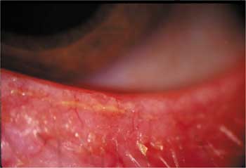
Inflammation of the eyelid margin is one of the classic signs of blepharitis. |
Curing the condition altogether is unlikely. Management is frustrating, since it is difficult to reduce the normal meibomian gland secretions. Maintenance therapy of warm compresses and lid cleansing with shampoo scrubs is crucial for success. In addition, patients should massage and express the glands regularly. When there is significant inflammation or suspected infection, prescribe an antibiotic ointment to be applied to the lids. Lens wear may be difficult until the condition is under reasonably good control.
Recently, some authorities have suggested using flax oil supplements (omega-3 fatty acid), either by pill or liquid, to stabilize the meibomian secretions. Some evidence suggests that the liquid variety is absorbed more effectively and produces a better result. If the patient chooses to use the pill form, prescribe an initial dose of 2000mg daily, tapered to 1000mg after a few weeks. Many contact lens wearers have reported improved tolerance to lens wear while taking flax oil.
It is imperative that soft lens wearers replace their lenses frequently because of the potential for excessive deposit buildup. Lens wearers often have vague symptoms of intolerance that initially outweigh the condition"s other clinical signs.
Seborrheic blepharitis with secondary meibomianitis is similar to meibomian seborrheic blepharitis, except that it produces sporadic episodes of inflammation. The secondary meibomianitis blocks the meibomian glands and anterior seborrhea; this manifests as stagnated or solidified meibomian secretions. The meibomian secretions have the consistency of toothpaste. The tear film is heavily contaminated and often shows significant debris and reduced quantity, but the bacterial flora is generally normal.
Treatment calls for conventional lid hygiene with massage and expression, and during exacerbations an antibiotic ointment applied to the lids. Perry and others have advised against using a detergent shampoo because of the potential for increased saponification (fats converted into soaps and glycerol) and contamination of the lipid layer that occurs in many patients. They advocate using a salt-water-only solvent when performing lid scrubs to avoid drying out the eyelid skin.
Also consider prescribing an oral tetracycline antibiotic. In resistant cases, we have used doxycycline 200mg daily for three to four weeks, followed by a maintenance dose of 50-100mg daily for several more weeks. Unlike in primary disease, you will rarely need to continue therapy for secondary meibomianitis beyond eight weeks; often the condition shows some improvement after just four to six weeks of treatment.
Withhold oral tetracyclines from children younger than eight and from women who are pregnant or nursing. In other patients, the major side effect is gastrointestinal distress. This is often dose-related; patients can ameliorate the distress by taking the medication with food. Over time, patients often begin to tolerate the medication better. Since these medications are considered photosensitizing agents, warn patients to avoid excessive sun exposure.
Primary Meibomianitis
Primary meibomianitis, or meibomitis, is a generalized sebaceous gland dysfunction commonly found in patients with seborrheic dermatitis or rosacea. It occurs more commonly in patients older than 50, and possibly in colder climates.
Cytologic studies show that the condition results from obstruction of the gland orifices by desquamated epithelial cells that tend to aggregate in keratotic clusters. Increased cellular debris thickens the gland"s lipids, leading to stagnated sebaceous secretions. This stagnation alters the oil layer"s contribution to the tear film.
The symptoms of primary meibomianitis mirror those of other forms of blepharitis. They include irritation, chronic burning, stinging, and foreign body sensation. Bacterial flora release free fatty acids from the meibomian lipids through the production of lipase and other hydrolytic enzymes. The free fatty acids can be toxic to the ocular surface, contaminating the tear film and producing significant inflammation.
| DEMODICOSIS |
|
Demodicosis is an inflammation of the lids attributed to a common mite that inhabits the follicle of the lash, especially in the elderly. Demodicosis is usually innocuous. Two different species produce varying effects. Demodex folliculorum leads to cuffing of the base of the cilia. D. brevis presents in the sebaceous, and in particular the meibomian glands. It has the potential to destroy the glandular cells, produce granulomas and plug meibomian glands. Some have postulated that this organism may be a cause for primary meibomianitis. Common symptoms are itching and burning, possible lid margin crusting and loss of lashes, along with the classic lash cuffing. Diagnosis is easy using simple epilation and examination with light microscopy to identify the mite. Application of cotton swabs saturated by ether and followed by antibiotic ointment is generally effective. Lens wear can resume with little or no interruption. |
In patients with rosacea, the cheek, nose, and forehead may have persistent erythema, telangiectasia, papules, pustules and hypertrophic sebaceous glands. In advanced cases, rhinophyma may occur. Signs of this condition include:
Inspissated plugs at the orifices of the meibomian glands.
Cloudy and thickened yellow-white secretions on gland expression.
Foamy discharge of the tear film meniscus.
Conjunctival hyperemia.
Thickened, rounded eyelid margins.
Often you may see multiple calcific concretions on the palpebral conjuctiva, along with signs of previ ously treated or untreated chalazia. Meibography through transillumination techniques shows the morphology of the glands with detail and is helpful in gauging the extent of involvement.
Contact lens evaluations of patients with primary meibomianitis reveal peculiar staining patterns and unstable, quick tear breakup times. Occasionally, peripheral corneal infiltrates and pannus may develop. Peripheral corneal vascularization may progress toward the visual axis with lipid deposition, scarring, and opacification at the leading edge. Visual impairment is always a possibility in neglected cases. Rarely, a peripheral ulcerative keratitis occurs with stromal thinning and possible perforation or microbial superinfection.
The most effective treatment for primary meibomianitis is warm compresses followed by lid massage and gland expression. Practitioners should perform these procedures in the office and then carefully instruct each patient on how to do them at home two to four times daily.
Oral tetracycline medications have become the main therapy for moderate to severe cases of meibomian gland dysfunction. Once you see that the initial dosage has been effective, you must keep the patient on a reduced long-term maintenance dose. For those who cannot tolerate oral tetracycline therapy, a safe and effective alternative is topical metronidazole gel 0.75 percent (MetroGel) applied to the eyelids.
 |
|
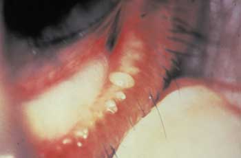
Meibomitis in a contact lens wearer. |
For dry eye that often accompanies this condition, the best management is artificial tears and lubricating ointments. Because patients must instill the tears frequently, it is best to prescribe a preservative-free or "disappearing-preservative" product. You can use silicone punctal plugs to relieve the patient"s symptoms, but only after you have addressed the stagnation effects on the tear film and the potential for infection by using lid hygiene techniques and perhaps an antibiotic ointment.
As with any form of blepharitis, be cautious in cases where you deem it appropriate to use a steroid or antibiotic-steroid combination. Topical steroids are useful in the first month of treatment to reduce ocular inflammation. In addition to the potential for side effects and over-use, some patients rely too heavily on this option rather than addressing the hygiene aspects of therapy. Compounded medroxyprogesterone 1% may be beneficial in cases involving progressive vascularization, corneal thinning or peripheral ulcerative keratitis.
Patients with primary meibomianitis--and especially those with accompanying rosacea--must curtail contact lens wear until the condition resolves to the point where simple lid hygiene offers adequate management. For most patients, this may take a long time. Many will not do well with contact lenses and must be advised of other options.
Angular Blepharitis
Angular blepharitis is a localized eczematoid inflammation of the lid at the outer canthus and sometimes medial canthal region. The staphylococcal form is typically dry and scaly. The Moraxella form is generally wet, and produces a macerated lid with a whitish, frothy discharge. Angular blepharitis may involve the conjunctiva as well. Clinicians have also identified gram-negative bacillus DF-2 as a possible cause of eczematoid blepharitis.
All forms of angular blephritis call for treatment with an antibiotic ointment. Once the condition has resolved--and ideally when the source of infection is identified--contact lens wear can resume.
Exercise Caution
Treating blepharitis centers on controlling the inflammation and preventing secondary ocular complications. Contact lens wear further complicates management. Lens wear may pose an added threat to the compromised ocular surface in patients with blepharitis. Symptoms of intolerance may worsen because of secondary tear film instability that accompanies many forms of blepharitis.
No complete cure exists for chronic blepharitis. A comprehensive strategy is vital to help patients who wish to wear contact lenses. This entails identifying and treating these conditions whenever possible before fitting a contact lens, and managing blepharitis aggressively when it develops in current lens wearers. Choosing the right material and replacement plan, setting an appropriate wearing schedule, and prescribing lubricants are essential measures to assure safe and effective lens wear. Advise your contact lens patients that they have additional risks for blepharitis, including increased surface deposits, increased inflammation and the potential for infection.
Dr. Shovlin is editor-in-chief of Review of Contact Lenses and associate clinical editor for Review of Optometry. He practices in Scranton, Pa. Dr. DePaolis is in private practice in Rochester, N.Y., where he"s on staff at the University of Rochester School of Medicine.
