 Multiple sclerosis (MS) is a chronic, recurrent disease characterized by disseminated patches of demyelination. Over time, patients manifest episodic neurologic dysfunction due to inflammation in different regions of the central nervous system.1 The most commonly affected areas are the brain, spinal cord and optic nerves—therefore, the optometrist may be the first physician to whom an MS patient presents.
Common neurologic symptoms of MS include sensory disturbances, motor weakness and trigeminal neuralgia. Patients may experience spontaneous and movement-induced muscle spasms, sensory problems (paresthesias and hypesthesia), ataxia, bladder dysfunction, constipation, cognitive dysfunction, depression, fatigue, sexual dysfunction, facial weakness, vertigo and hearing loss.2
In the first part of this two-part column (“
MS and the Eye (Part 1),” January 2012), we presented an overview of demyelinating disease, with a focus on MS. In Part 2, we discuss the classification, diagnosis and management of MS.
Common Neuro-ophthalmic Symptoms
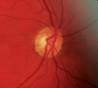
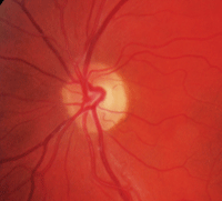
Disc photos of a 58-year-old white female with long-standing multiple sclerosis. Note areas of optic nerve atrophy and nerve fiber layer thinning.
Common neuro-ophthalmologic symptoms in MS are unilateral vision loss due to optic neuritis, oscillopsia due to nystagmus, and diplopia due to internuclear ophthalmoplegia (INO) or cranial neuropathy. The occurrence of bilateral INO is considered to be highly suggestive of MS, especially in young patients. A new-onset acquired pendular nystagmus is relatively common, but other forms of nystagmus may occur as well, depending on the location of the demyelinating lesion.
Combinations of deficits, including horizontal or vertical gaze palsies, wall-eyed bilateral INO or wall-eyed monocular INO, or paralytic pontine exotropia (the “one-and-a-half syndrome”), may also occur in MS.3 (See “ Common Ophthalmic Manifestations of MS,” below.)
Classification System
MS has four clinical types:2-4
• Relapsing/remitting (RRMS) accounts for 85% of MS cases at onset. Attacks evolve over days to weeks, but there is usually complete recovery over the ensuing weeks to months. Between attacks, patients are neurologically stable.
• Secondary progressive MS (SPMS) begins as RRMS and then changes during the clinical course so that the patient experiences steady deterioration in function that isn’t associated with acute attacks. SPMS produces a greater amount of fixed neurologic disability than RRMS.
• Primary progressive MS (PPMS) patients do not experience attacks, only a steady decline from the disease onset.
• Progressive relapsing MS (PRMS) patients experience steady deterioration in their condition from the onset; however, as in SPMS, there are occasional attacks that overlap with the progressive course.
Establishing a Diagnosis
An MRI scan (T1 and T2) with fluid attenuated inversion recovery sequencing (FLAIR) and gadolinium infusion is the neuroimaging study of choice. MRI is excellent for identification of white-matter plaques, and is superior to CT scan for visualizing the posterior fossa and spinal cord. Plaque lesions may take on a spherical or ovoid configuration, and vary in size from 1mm or 2mm to several centimeters. Plaques are often situated at the ventricular margins, optic nerves and chiasm, corpus callosum, spinal cord, and throughout the brain stem and cerebellar peduncles.2-4
The cerebrospinal fluid (CSF) in patients with definite MS is abnormal in more than 90% of cases. The most common abnormalities are increased levels of immunoglobulin G (IgG), the elevation of the IgG/albumin index and the presence of oligoclonal IgG bands.3 There are no definitive diagnostic tests for MS, so the disease remains a clinical diagnosis.1-3 The McDonald criteria for diagnosing MS allows for the use of paraclinical data, which includes MRI and CSF studies.5
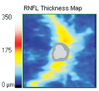
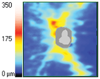
Advances in ophthalmic imaging with OCT have made it possible to quantify RNFL atrophy as a structural marker of axonal injury in the afferent visual pathway in patients with MS.
Advances in ophthalmic imaging with optical coherence tomography (OCT) have made it possible to quantify retinal nerve fiber layer (RNFL) atrophy as a structural marker of axonal injury in the afferent visual pathway in patients with MS. Studies have shown that OCT-measured RFNL values are reduced in patients with MS with and without a history of optic neuritis; however, RNFL atrophy tends to be greater in eyes affected by optic neuritis.6
Therapeutic Options and Considerations
There is no cure for MS. The early promotion of remyelination and preservation of oligodendrocytes remain important therapeutic goals.1-3
Common
Ophthalmic Manifestations of MS
• Optic neuritis—inflammation of optic nerve in one or both eyes, typically retrobulbar.
• Nystagmus—typically pendular, but upbeat, downbeat, convergence-retraction may also occur.
• Internuclear ophthalmoplegia (INO)/bilateral INO—adduction deficit of ipsilateral eye with horizontal gaze nystagmus in the contralateral abducting eye.
• Cranial nerve palsies (III, VI)
Therapy for MS can be divided into several categories: treatment of acute attacks as they occur; treatment with disease-modifying agents that reduce the biological activity of MS; and symptomatic therapy.1
High-dose, intravenous corticosteroids (methylprednisolone 500mg/d to 1,000mg/d for three to five days, followed by a course of oral prednisone beginning at a dose of 60mg/d to 80 mg/d and tapered over two weeks) are often used to treat acute exacerbations of MS. However, no evidence shows lasting improvement or alteration of prognosis with corticosteroid administration.
Other immunosuppressive and immunomodulating agents—Avonex (interferon beta-1a, Biogen Idec.), Rebif (interferon beta-1a, EMD Serono, Inc. and Pfizer Inc.), Betaseron (interferon beta-1b, Bayer Healthcare), Copaxone (Glatiramer, Teva) and Tysabri (natazilumab, Biogen Idec and Elan Pharmaceuticals, Inc.)—may be useful in long-term treatment.1,3 Current research suggests that early diagnosis and therapy is critical.3
Mortality as a direct consequence of MS is uncommon. Death can occur during an acute MS attack; although this is a rare event. More commonly, death occurs as a complication of MS (e.g., pneumonia).1
Differential Diagnosis
It is important to note that neuromyelitis optica (NMO), another idiopathic inflammatory demyelinating disease of the central nervous system, also predominantly affects the optic nerves and spinal cord and should be highly considered as a differential diagnosis to MS. NMO antibodies (aquaporin-4 antibodies) can be routinely tested and have become a major diagnostic criteria for NMO. However, there is still some debate among the neuro-ophthalmology community if the aquaporin-4 antibodies should be routinely tested in patients with isolated optic neuritis.
Demyelinating disorders are characterized by inflammation and selective destruction of the central nervous system myelin.1 Because MS is one of the most common acquired neurologic disorders in the United States, it is important for optometrists to be familiar with both its ocular and neurologic consequences so that they can make an accurate diagnosis and administer proper management.2,3
Who Gets MS?7
• MS is at least two to three times more common in women than men.
• Most people are diagnosed between the ages of 20 and 50; however, MS
can appear in young children and teens as well as much older adults.
• MS occurs in most ethnic groups, including African-Americans, Asians
and Hispanics/Latinos, but is more common in Caucasians of northern
European ancestry.
1. Hauser SL, Goodin DS. Multiple sclerosis and other demyelinating diseases. In: Hauser SL, Josephson SA. Harrison’s Neurology in Clinical Medicine. 2nd ed. New York: McGraw-Hill; 2010:435-50.
2. Motor deficits. In: Simon RP, Greenberg DA, Aminoff MJ. Clinical Neurology. 7th ed. New York: McGraw-Hill; 2009:152-202.
3. The M.D. Association. Selected systemic conditions with neuro-ophthalmic signs. In: American Academy of Ophthalmology. Basic and clinical science course, section 5: neuro-ophthalmology; 2006-2007:317-68.
4. Introduction, organization, and cellular components. In: Young PA, Young PH, Tolbert DL. Basic Clinical Neuroscience. 2nd ed. Philadelphia: Lippincott Williams & Wilkins; 2008:1-14.
5. McDonald WI, Compston A, Edan G, et al. Recommended diagnostic criteria for multiple sclerosis: guidelines from the International Panel on the diagnosis of multiple sclerosis. Ann Neurol. 2001 Jul;50(1):121-7. Table 3.
6. Lamirel C, Newman N, Biousse V. The use of optical coherence tomography in neurology. Rev Neurol Dis. 2009 Fall;6(4):E105-20.
7. National Mutiple Sclerosis Society. Available from:
www.nationalmssociety.org (accessed Feb 14, 2012).
