Whether you practice in Portland, Maine, Portland, Ore., or some location in between, a new report indicates you’ll have more patients sitting in your chair with diabetic retinopathy than ever before.
The report, Vision Problems in the U.S., by Prevent Blindness America and the National Eye Institute (
www.visionproblemsus.org), cites an 89%spike in diabetic eye disease from 2000 to 2010, based on U.S.Census data and 12 epidemiological studies. The report, prepared by researchers at John Hopkins University, indicates that 7.69 million people in the U.S. age 40 and older suffer from diabetic retinopathy, up from 4.06 million in 2000. Additionally, those in the 40-plus age bracket with vision impairment and blindness from any etiology has increased by 23% since 2000.

“Rates of diabetes are growing alarmingly, so it’s not surprising that cases of diabetic retinopathy have gone up,” says Washington State optometrist and diabetes specialist Paul Chous, O.D., who has had type 1 diabetes for 44 years.
Troubling Statistics

The findings may not be a surprise, but the escalating diabetic retinopathy rates are still troubling.
“We are so fortunate to have access to information like this from Prevent Blindness America and NEI; however, I am disheartened by the data,” says optometrist and certified diabetes educator Tina MacDonald, O.D., of Los Angeles.“Diabeticretinopathy has increased by more than 89%, while those in the United States population40 and older grew by 19.5% between 2000 and 2010—it’s really a shocking number.”
Prevent Blindness America recently unveiled the Vision Problems in the U.S. report during the “Focus on Eye Health” summit in Washington, D.C., where the Centers for Disease Control and Prevention’s Division of Diabetic Translation presented some alarming statistics:
- 26 million Americans suffer from diabetes and 79 million more have pre-diabetes.
- One in three U.S. adults could have diabetes by 2050 if current trends continue.
- Diabetic retinopathy is the leading cause of new cases of legal blindness among adults ages 20 to 74 in the U.S.
“Notably, there are seven million people who are undiagnosed,” says Jinan Saaddine, M.D., medical epidemiologist at the CDC’s Division of Diabetes Translation and leader of its Vision Health Initiative. Dr. Saaddine attributes the burgeoning diabetes rate to several factors, including an aging population, growing trends of obesity and sedentary lifestyles, and an increase in minority populations that are at high risk for developing diabetes.
Taking a closer look at the data reveals emerging trends about which patients are most affected now as well as who’ll be at high risk in the future.
The Nuts and Bolts of the Vision Problems in the U.S. Report
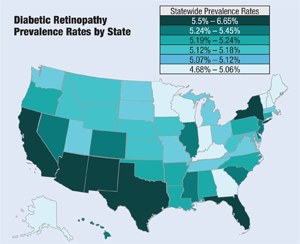
- The study found states with the highest prevalence of diabetic retinopathy, in order, are New Mexico (6.6%), Florida (6.0%), Texas (5.9%), California (5.8%), Arizona (5.8%), the District of Columbia (5.5%) and New York (5.5%). Why these particular states? Dr. Varma attributes it to a higher Hispanic and aging population in the majority of these states. Interestingly, however, the CDC reports that counties and states in the southeast region of the country have the highest rates of diabetes, according to Dr. Saaddine.
- The age group with the highest prevalence of diabetic retinopathy by percent (among both men and women) is 65 to 74. The age group with the highest number of cases is 50 to 64 (3.2 million).
- Data from this report was derived from pooling information from 12 major epidemiological studies and accounting for the number of individuals with diabetic retinopathy, the number at risk, age, race and gender. U.S. Census 2010 data was then applied to generate the final prevalence rates in the various categories.
- The numbers reported in this study are estimates, not exact measurements. All data from the report can be obtained through a new searchable database on the Prevent Blindness America website: www.preventblindness.org/visionproblems. This tool enables users to research a wide range of information, including eye disease and condition numbers broken down by state, age, gender and race, and provides comparisons across disease conditions.
DR in Hispanics
Hispanics, already one of the largest ethnic groups in the U.S., accounted for more than half the growth in the total population between 2000 and 2010, according to the latest census. This group added 15.2 million people during the period studied, which constitutes about 56% of the total U.S. population growth of 27.3 million.
Results from the Vision Problems in the U.S. report show that Latinos/Hispanics had the highest rate of diabetic retinopathy (8%) compared to whites (5%), blacks (5%) or other groups (4%). With disease prevalence for all U.S. citizens age 40 and older at 5.4%, only Latinos exceeded the national average.
This comes as no surprise to Rohit Varma, M.D., M.P.H., principal investigator of the Los Angeles Latino Eye Study.1 Dr. Varma’s seminal study found that Latinos have higher rates of developing visual impairment, blindness, diabetic eye disease and cataracts than non-Hispanic whites. “We know for a fact that Latinos have one of the highest prevalence rates of type 2 diabetes in the U.S.,” Dr. Varma says. “And, the overall rate of Latinos who have diabetic retinopathy is on the higher end than non-Hispanic whites.”

Studies are finding a high prevalence of diabetic eye disease among Hispanics because this ethnic group has a greater genetic predisposition to retinopathy than non-Hispanic whites, Dr. Varma says. “My belief is that their risk of developing retinopathy is higher at the same level of the risk
factor. For example, Hispanics have a greater rate of developing eye disease than others with same blood pressurelevel.” Dr. Varma says this is primarily due to a greater genetic predisposition in Hispanics.

In addition to a predisposition to develop diabetes, socioeconomic factors may play a part in higher prevalence rates of diabetic retinopathy in certain ethnicities, such as Latinos and blacks, Dr. Chous says. These factors include lack of access to high-quality health care and even grocery stores, he adds.
The report’s finding that blacks had the same diabetic retinopathy prevalence rate as whites came as a surprise, Dr. Chous says, as previous reports indicate that blacks are about 50% more likely to develop retinopathy, compared to their European-American counterparts.2 This statistic has been attributed to poorer health care access and higher rates of hypertension in the former community.2
“If this extrapolated statistic is true, this new finding may reflect the success of increased public and community education efforts developed within and aimed at the African-American community,” Dr. Chous says.
However, it could also be related to higher rates of diabetic retinopathy among all Americans with diabetes in a bad economy where the financial costs of good self-care can take a back seat to other financial demands, he adds. “This is not speculation. I saw a 25-year-old unemployed, type 1 Caucasian paramedic earlier this year who just lost his vision to proliferative retinopathy after taking his insulin injections every other day to save money,” Dr. Chous says.
According
to the Vision Problems in the U.S. report, New Mexico is the state with
the highest prevalence of diabetic retinopathy in the nation (6.6%).
For Brent Shelley, O.D., who practices in La Mesilla, N.M., this is no
surprise. “I would say that it
corresponds with the rates seen in our practice,” says Dr. Shelley,
president of the New Mexico Optometric Association. “Our state has a
high percentage of Hispanics and Native Americans, and these
ethnicities, in turn, have high incidences of diabetes. Furthermore, our
state is very rural by nature and primary care, in general, is
underserved. Hence, outreach programs are very much needed so that we
can educate these persons to reverse this statistic.” While
the rates of diabetes and diabetic retinopathy are increasing, Dr.
Shelley says he has not seen an increase in sight-threatening cases.
“However, I will say that early diabetic retinopathy is becoming more
frequent,” he says. Because Dr.
Shelley’s practice is located in a medical plaza, he receives many
primary care referrals. “We educate our patients thoroughly, maintain an
aggressive recall strategy and correspond with the patient’s physician
regarding their ocular health,” he says.
For
Dr. Shelley, the optometrist’s standard of care in a diabetic patient
is to dilate and educate the patient on the pathophysiology of diabetes.
“In our practice, we will not see a diabetic patient without dilating
their eyes. While some practices may rely on fundus imaging for retinal
exams, I would argue that this does not meet the standard of care and is
not in the patient’s best interests,” he says.
Treating Diabetic Retinopathy in New Mexico
Keeping Statistics in Mind
The 89% spike in diabetic retinopathy cases as detailed by the Vision Problems in the U.S. report is a bit misleading, as this is not a medical study but rather a demographic statistical report, says Ojai, Calif. optometrist Roger Phelps, one of the few O.D.s who is also a certified diabetes educator. Dr. Phelps has had type 1 diabetes for more than 20 years. “The 89% figure mainly reflects the increasing number of people with diabetes in our country, not a disease process that is getting out of control,” says Dr. Phelps. “The opposite is actually true: Although we have many more patients with diabetes, these individuals can be offered a plan to greatly reduce their risk of vision loss from diabetes.”
All the studies Dr. Phelps has reviewed agree with the report relative to the increasing number of people in the country with diabetes, due to population increases—especially in the Baby Boomer generation—as well as an increasing percentage of the population who have diabetes.
However, many studies show those individuals with diabetes are much less likely to lose vision now than 15 years ago with proper education and care, he adds. Dr. Phelps cites the Wisconsin Epidemiology Study of Diabetic Retinopathy, which found the lower risk of proliferative diabetic retinopathy in more recently diagnosed patients possibly reflects improvement of care.3
Dr. Chous offers another possibility for the high prevalence of diabetic retinopathy: Because diabetic retinopathy detection has increased as a function of improved technology (e.g., greater use of retinal imaging with red-free filters, widefield imaging and OCT), rates of actual disease (per 1,000 people diagnosed with diabetes, for example) may not have grown as much as it would appear.
While the 89% figure certainly is significant, it is also critical to distinguish between nonspecific diabetic retinopathy (mild to severe) and sight-threatening diabetic retinopathy (proliferative diabetic retinopathy and clinically significant macular edema) that causes visual disability, Dr. Chous says, as most cases of diabetic retinopathy do not lead to vision loss.
From 2005 to 2008, 4.2 million (28.5%) people with diabetes age 40 or older had diabetic retinopathy; of these, 655,000 (4.4% of those with diabetes) had advanced diabetic retinopathy that could lead to severe vision loss.2
“Also, every retinal specialist will tell you that rates of severe vision loss from diabetic retinopathy have gone down with improvements in diabetes care—especially blood glucose—improved detection and earlier diagnosis, and better treatments to prevent vision loss,” says Dr. Chous.
However, even mild diabetic retinopathy has been linked to increased risk of other diabetes complications, including kidney and cardiovascular disease, so from a public health perspective, increased rates or detection of diabetic retinopathy are important and underscore the need to prevent the onset of diabetes, Dr. Chous adds.
The Optometrist’s Role in DR
As studies such as the Vision Problems in the U.S. report find that more patients are developing diabetes––and diabetic retinopathy as a result––the optometrist’s role in treating diabetes patients becomes more critical than ever.
“We can encourage our patients to get to know their disease, and as they do, they can fully participate in their care and do much to stop the loss of vision that was so prevalent in the past,” Dr. Phelps says. He recommends optometrists implement the following in caring patients with diabetes:
• Know your patient’s A1C level, and encourage patients to know it as well. (See the “The ABCs of A1Cs.") If a patient’s A1Cs are staying significantly over 7%, let them know that they are at a higher risk of retinopathy even though there may not be any diabetic changes in their eyes today. Encourage them to discuss and agree upon the appropriate A1C level for their individual medical status with their primary care physician. When a patient who returns for an annual dilated exam shows increasing retinopathy, it is time to move the slit lamp aside and have another discussion about glycemic control. Realize that, even with an improved A1C, ocular changes can take place up to three years later from prior poor control.4 If a patient’s A1C is still above the goal set with their physician and increasing retinopathy is present, it may be best to get a retinal consult even before pre-proliferative changes are apparent.
• Perform a dilated exam annually. “We may find many patients with non-threatening mild non-proliferative retinopathy, but let the patient know of the importance of regular eye exams, as we can also spot asymptomatic sight-threatening changes,” Dr. Phelps says.
Diagnostic Codes for DR
• Always send a report of your retinal findings to the patient’s primary care physician after the exam, even if there are no signs of diabetic retinopathy. This report can simply state: “No diabetic retinopathy.”
• Consider an OCT if a patient is developing even moderate levels of diabetic retinopathy. “I recommend an OCT sooner rather than later, because sometimes it might show changes you might not normally see, and it will provide a baseline,” Dr. Phelps says. “When I see a few minor changes in the retina such as microaneurysms and small blot hemorrhages, it is mild. When more of these are present in more than three quadrants combined with some cotton wool spots, it gets into the moderate range,” Dr. Phelps explains.
• Work closely with retinal specialists in your area and make timely referrals.
“Although we have a confirmed epidemic increase in our national patient population of those with diabetes, we have made major progress in individual prevention of vision loss,” Dr. Phelps says. Most vision loss from diabetes has now been shown to be preventable by teaching patients the importance of controlling their diabetes (as best reflected in the A1C testing two to four times a year), and for them to return each year for a dilated eye exam to discern the need for timely laser treatment or anti-VEGF injections for DME, he adds.
Patients who have suffered vision loss as a result of diabetic retinopathy also need low vision rehabilitation, in addition to managing their diabetes, Dr. MacDonald adds.
“Optometrists are really good at educating patients, but we are going to have to do even better,” Dr. MacDonald says. “Diabetic retinopathy is the leading cause of blindness among those of working age, and it doesn’t have to be that way.” With appropriate treatment, she says, progression can be stopped. “There are health disparities among racial and ethnic groups that we need to be aware of. We need to get people in the habit of, and emphasize the need for, complete dilated exams even if there are no symptoms. Finally, we need to have good communication not only with the patient, but also the rest of the health care team.”
As the diabetic
population in the U.S. continues to grow, Dr. Phelps recommends that
every optometrist should at least know and document their diabetic
patients’ A1C levels. A1C is a
blood test that gives the average amount of glucose in the blood over
the past three to four months. An A1C of 5.6% or below is normal. In
pre-diabetes, A1C levels range between 5.7% and 6.4%. If the A1C is 6.5%
or above, the patient has diabetes. Also
be aware that if your patient’s A1C level increases, there generally is
a three-year time delay until it will impact their eyes. Refractive
shifts can show up quickly with a rapid A1C change as well, Dr. Phelps
says. “If a person comes in,
and their A1C had been maintained at around 7% but now it’s up to 12%,
it may be two to three years until it affects their eyes, but when it
does, there is very little you can do to get it back,” Dr. Phelps says.
“If the A1C levels are truly going up, even if there is no diabetic
retinopathy, I am still very concerned.” Another
critical factor to keep in mind: In general, every percentage point
drop in A1C blood test results (e.g., from 8% to 7%) can reduce the risk
of microvascular complications (eye, kidney and nerve diseases) by
40%.5
“Optometry needs to
learn the importance of monitoring their patients’ A1Cs. If a patient
doesn’t know what their level is, you need to ask them: ‘May I call your
doctor?’” Dr. Phelps says.
Optometrists
should encourage their patients with diabetes to see their primary care
physician regularly so their doctor can monitor them regularly and set
their A1C goals. They should also get to know the physicians in their
area and personally call their patients’ primary care doctors, to find
out A1C levels, Dr. Phelps advises.
The
A1C level may affect the glasses you prescribe now, as well as their
risk of retinopathy in the future. You can let the M.D. know that you
just saw your common patient, and although the patient’s eyes show no
diabetic retinopathy (or level of retinopathy), you want to know the
patient’s current A1C or recent history of A1C. If the patient’s A1C is
variable, recheck the refraction in a few weeks. If the A1C has been
stable over the past three to six months, you can prescribe glasses
right away, Dr. Phelps says. Developing
a dialog with the patient’s primary care physician can help the
optometrist become a “team member” in the care of their mutual patient,
Dr. Phelps adds.
The ABCs of A1Cs
1. Varma R, Ying-Lai M, Klein R, Azen SP. Prevalence and risk indicators of visual impairment and blindness in Latinos: The Los Angeles Latino Eye Study. Ophthalmology. 2004 Jun;111(5):1132-40.
2. Zhang X, Saaddine JB, Chou CF, et al. Prevalence of diabetic retinopathy in the United States, 2005-2008. JAMA. 2010 Aug 11;304(6):649-56.
3. Klein R, Knudtson MD, Lee KE, et al. The Wisconsin Epidemiologic Study of Diabetic Retinopathy: XXII the twenty-five-year progression of retinopathy in persons with type 1 diabetes. Ophthalmology. 2008 Nov;115(11):1859-68.
4. The effect of intensive treatment of diabetes on the development and progression of long-term complications in insulin-dependent diabetes mellitus. The Diabetes Control and Complications Trial Research Group. N Engl J Med. 1993 Sep 30;329(14):977-86.
5. Centers for
Disease Control and Prevention. National diabetes fact sheet: national
estimates and general information on diabetes and prediabetes in the
United States, 2011. Atlanta. U.S. Department of Health and Human
Services, Centers for Disease.
