There are eight distinct DNA viruses that infect humans, and five of them cause significant ocular disease. These include: HSV-1, HSV-2, varicella-zoster, cytomegalovirus and Epstein-Barr virus.1

HSV-1, which most commonly affects the eyes, is almost universally acquired. In fact, by age 60, more than 90% of individuals have HSV-1 in their trigeminal ganglia.2 To date, more than 500,000 Americans have been diagnosed with HSV ocular infections, with about 20,000 new cases and 28,000 recurrences reported each year.3
The breakdown is interesting, with 72% of HSV-1 cases involving the epithelium, 41% involving the lids and conjunctiva, 12% the corneal stroma and 9% involving the uveal tract. It’s interesting to note that humans are the only natural reservoir of HSV and that the virus can remain viable for up to two hours on a dry tonometer head or more than eight hours on a wet tonometer tip.4
Here, we review the ophthalmic ramifications of HSV infection as well as discuss some of the latest treatment options.
Primary HSV
- Overview. Typically, HSV-1 infection occurs relatively early in life; however, recent research suggests that primary acquisition of HSV-1 is becoming progressively delayed in industrial countries.2 In most cases, patients with primary HSV exhibit mild flu-like symptoms that may be associated with a low-grade fever. A small percentage of individuals also experience viral skin eruptions, which generally manifest as a vesicular rash found around the eyelids, conjunctiva or facial skin. Often, pre-auricular node swelling and pain also accompany these skin eruptions. After the primary infection runs its course, the HSV becomes latent in the trigeminal ganglion. Recurrent HSV typically occurs in the oral or nasal mucosa because of certain reactivation triggers, such as local trauma from ocular surgery, UV light exposure, hormonal changes, stress and systemic illnesses that compromise the immune system.5
- Treatment. Children who manifest primary HSV usually are less responsive to medications, although they still should be treated with anti-viral medications, such as oral acyclovir or topical antivirals (if a conjunctivitis is present). Topical corticosteroids should be avoided because of the risks associated with further immune system suppression. It is also important to note that laser surgery procedures, such as LASIK and PRK, can reactivate a latent HSV virus.6,7 Additionally, corticosteroids have not been shown to be a trigger for HSV reactivation. Nevertheless, if a reactivation occurs in the presence of steroids, the disease can progress aggressively.8
A Look at Ocular HSV
Corneal involvement of HSV can include epithelial keratitis, neurotrophic keratopathy, stromal keratitis and endotheliitis. The infectious epithelial keratitis presentation includes corneal vesicles, dendritic keratitis and geographic ulcers. Corneal vesicles are the corollary to the vesicular lesions found on the skin or eyelids. They are cystic or small, raised, clear lesions that contain the active virus. Corneal dyes are an important diagnostic tool in HSV identification (although epithelial defects will not be apparent at this stage, you will note inverse or negative staining or pooling of the fluoresceine around the raised vesicles). This is a rare clinical presentation because patients typically are not very symptomatic, but as the vesicles coalesce, they form the classic dendritic ulcer presentation.
The dendritic ulcer is the most common presentation of ocular HSV. The features include a branching lesion with swollen epithelial borders. It is sometimes difficult to differentiate a pseudodendrite (i.e., as seen in herpes zoster) from HSV. The key is to realize that HSV dendrites are true ulcers, so they accrue stain within the lesion as well as along the borders and terminal end bulbs. This can often be enhanced with the addition of lissamine or rose bengal dye.
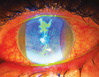
New treatments for ocular herpes could potentially benefit this patient who presented with significant iritis secondary to HSV.
The geographic ulcer is essentially an enlarged or expanding dendritic ulcer that often presents with scalloped borders. The geographic ulcer can occur if a patient with a dendritic ulcer is placed on a corticosteroid. Other forms of HSV keratitis include the marginal ulcer. This lesion mimics a Staphylococcal hypersensitivity or sterile infiltrative keratitis as a lesion near the limbus. Because the location is in close proximity to the blood vessels, it causes rapid infiltration of white blood cells under the ulcered area. Patients with HSV marginal ulcers are more symptomatic than patients with sterile limbal infiltrates. Additionally, HSV marginal ulcers do not exhibit a clear zone between the infiltrate and the limbus, and they show the presence of deep stromal vessels.
Research shows that 10% of patients with epithelial keratitis go on to experience stromal disease within one year.9 Stromal forms of keratitis include neurotrophic keratopathy, immune stromal keratitis (ISK) or interstitial keratitis and endotheliitis. Stromal haze and neovascularization (also known as ghost vessels) with an intact epithelium are the hallmark signs of ISK. Occasionally, it can present as an immune ring. This presentation is most common in recurrent HSV disease and may often be accompanied by cell and flare in the anterior chamber.
Finally, disciform endotheliitis presents with a deep disc-shaped area of edema and keratic precipitates on the endothelium but no stromal infiltrate or neovascularization. Typically, endotheliitis has a corresponding mild to moderate iritis and is often associated with elevated IOP.
New Treatment Options
During the last decade, there has been a trend toward the use of oral antivirals for the treatment of all forms of HSV. This shift was due, in part, to higher rates of corneal toxicity that were associated with the topical antiviral medications that were available 10 years ago. It is interesting to note that trifluridine was approved in the U.S. more than 30 years ago, and only last year did we see the introduction of a new topical antiviral––Zirgan (0.15% ganciclovir gel, Bausch + Lomb).
- Zirgan. This prodrug is only activated by the viral enzyme thymidine kinase to inhibit synthesis of viral DNA. Because of this, Zirgan is more selective than other antiviral agents and does not target healthy corneal cells. Zirgan should be dosed five times a day until the dendritic ulcer is healed, and then three times a day for an additional seven days. It does not require refrigeration and unlike Viroptic (trifluridine, Monarch Pharmaceuticals), Zirgan is preserved with 0.075mg of BAK. It also has a comparable pH and osmolarity to natural tears.
- Valtrex. Another development in the improved management of HSV is the increased availability of oral Valtrex (valacyclovir, GlaxoSmithKline), which became available as a generic medication in November 2009. Although all oral antivirals demonstrate low toxicity and are very effective, valacyclovir has five times the bioavailability of acyclovir.10
Still, with the approval of Zirgan and its selective antiviral properties and lessening of epithelial toxicity, we may again see clinicians switching back to topical medications for managing HSV keratitis and limiting systemic drug exposure.
New developments in HSV keratitis treatments have prompted clinicians to stay abreast of the most effective management strategies of this multi-presentation virus. Understanding the various forms of HSV keratitis can aid in accurate diagnosis and successful treatment, thus reducing the risk for associated corneal morbidity.
Dr. Karpecki is a paid consultant to Bausch + Lomb. Neither he nor Dr. Shechtman have any direct financial interest in the products mentioned.
1. Liesegang TJ.Herpes simplex virus epidemiology and ocular importance. Cornea. 2001 Jan;20(1):1-13.
2. Liedtke W, Opalka B, Zimmermann CW, Lignitz E. Age distribution of latent herpes simplex virus 1 and varicella-zoster virus genome in human nervous tissue. J Neurol Sci. 1993 May;116(1):6-11.
3. Young RC, Hodge DO, Liesegang TJ, Baratz KH. Incidence, recurrence and outcomes of herpes simplex virus eye disease in Olmsted County, MN. 1976-2007: The effect of oral antiviral prophylaxis. Arch Ophthalmol. 2010 Sep;128(9):1178-83.
4. Nagington J, Sutehall GM, Whipp P. Tonometer disinfection and viruses. Br J Ophthalmol. 1983 Oct;67(10):674-6.
5. Toma HS, Murina AT, Areaux RG Jr, et al. Ocular HSV-1 latency reactivation and recurrent disease. Semin Ophthalmol. 2008 Jul-Aug;23(4):249-73.
6. Asbell PA. Valacyclovir for the prevention of recurrent herpes simplex virus eye disease after excimer laser photokeratectomy. Tr Am Ophth. 2000;98:285-303.
7. Levy J, Lapid-Gortzak R, Klemperer I, et al. Herpes simplex virus keratitis after laser in situ keratomileusis. J Refract Surg. 2005 July-Aug;21(4):400-2.
8. Kibrick S, Takahashi G, Liebowitz H, Laibson PR. Local corticosteroid therapy and reactivation of herpetic keratitis. Arch Ophthalmol. 1971 Dec;86(6):694-8.
9. The Herpetic Eye Disease Study Group. A controlled trial of oral acyclovir for the prevention of stromal keratitis or iritis in patients with herpes simplex virus epithelial keratitis. Arch Ophthalmol. 1997 Jun;115(6):703-12.
10. Weller S, Blum M, Smiley M. Phase I pharmacokinetics of the acyclovir prodrug, valacyclovir. Antiviral Res. 1993;20(Suppl 1):144.
