A 60-year-old Hispanic female presented for an annual evaluation. The patient reported that her vision was slightly blurred in both eyes. Additionally, she noted that her eyes felt dry.
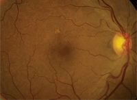
1. Fundus image of the posterior pole and macula of our patient's right eye.
Her medical history was significant for hypertension and rheumatoid arthritis. She was taking several medications, including an antihypertensive agent and 200mg Plaquenil (hydroxychloroquine, Sanofi-Aventis) b.i.d. The patient reported that she had been taking Plaquenil for the past two years. Her ocular history was unremarkable.
On examination, her best-corrected visual acuity measured 20/20 O.U. Her pupils were equally round and reactive to light, with no afferent defect. Confrontation visual fields were full to careful finger counting O.U., and ocular motility testing was normal.
Amsler grid testing was normal O.U. Color vision testing was normal (15/15 plates were correct). The anterior segment examination was remarkable for a scanty tear film and mild punctate epithelial erosions. Her intraocular pressure measured 15mm Hg O.U.
On dilated fundus examination, her optic nerves appeared healthy with a small cup and good rim coloration and perfusion O.U. The vessels were of normal caliber, and her peripheral retinae were unremarkable. Of interest, we noted changes that were located slightly superior to the right macula (figure 1). The left eye was remarkable only for a few drusen. We also performed a spectral-domain optical coherence tomography (SD-OCT) scan (figure 2).
Take the Retina Quiz
1. Which test was truly unnecessary to perform?
a. SD-OCT.
b. Color vision.
c. Amsler grid.
d. Both B and C.
2. What additional testing is necessary to appropriately manage this patient?
a. 10-2 visual field.
b. Pattern electroretinogram (ERG).
c. Fundus autofluorescence (FAF).
d. Electro-oculogram.
3. What does the SD-OCT scan reveal?
a. Normal anatomy.
b. Loss of the photoreceptor integrity layer (PIL).
c. Mild cystoid macular edema (CME).
d. Choroidal neovascular membrane (CNV).
4. What do the findings in the right macula represent?
a. Old branch retinal vein occlusion (BRVO).
b. Hydroxychloroquine toxicity.
c. Wet age-related macular degeneration.
d. Macular telangiectasia.
5. How should we manage our patient?
a. Recommend discontinuation of Plaquenil.
b. Close observation.
c. Anti-vascular endothelial growth factor (VEGF) therapy.
d. Anti-VEGF therapy and discontinuation of Plaquenil.
For answers, see below.
Discussion
There are several small microaneurysms located slightly superior to the right macula. The microaneurysms are seen as small, red bulbs with some fine telangiectatic vessels. Additionally, there are some retinal veins that are sheathed or perhaps even sclerosed. Could this be the result of Plaquenil use?
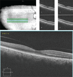
The SD-OCT scan of our patient's right eye. What do you notice?
Retinal toxicity caused by Plaquenil is quite rare considering the large number of people who use this drug. Nonetheless, clinicians (and most patients) are keenly aware of the potential ocular complications of this medication.
Patients who present with Plaquenil toxicity typically exhibit depigmentation that is located circumferentially around the macula, giving it a “bull’s eye” pattern. Consequently, this clinical finding is often termed bull’s eye maculopathy.
Bull’s eye maculopathy will yield the appearance of a ring-shaped scotoma on threshold visual field testing. This explains why patients on Plaquenil therapy should be followed with 10-2 visual fields. Unfortunately, once these changes are seen, the toxicity may already be fairly advanced.
For years, the single greatest problem that clinicians have faced is an inability to detect associated retinal changes earlier. But, with the advent of SD-OCT and other specialized ancillary tests (e.g., FAF and pattern ERG), these changes can be detected significantly earlier even before they are seen funduscopically and perhaps before they ever appear on visual field testing.
In early 2011, Michael Marmor, M.D., and colleagues published revised recommendations for screening patients who are on Plaquenil therapy.1 While the older recommendations focused on the maximum daily dose for a patient, the revised guidelines emphasized cumulative dose as the most critical factor for developing associated retinal toxicity.2,3
The traditional protocol for following patients on Plaquenil therapy consisted of baseline photography as well as annual color vision testing, Amsler grid and 10-2 visual fields annually.2,3 Under the new guidelines, color vision and Amsler grid testing are no longer considered acceptable screening methods so they have been removed from the protocol.2,3 However, 10-2 visual fields are still important. In addition, the authors recommended that visual fields be supplemented with any one of the following tests: multifocal ERG, SD-OCT or FAF.1 (The authors specifically noted that time-domain OCT was not sensitive enough to detect early changes.1)
Dr. Marmor’s research team also recommended that screening should be performed within the first year of initiating therapy, and then annually after five years of Plaquenil use. The risk for retinal toxicity begins once the patient has used Plaquenil for five to seven years and/or has taken a cumulative dose of more than 1,000g. Patients with renal or hepatic dysfunction are at an increased risk of toxicity.1 Other risk factors for retinal toxicity include short stature, obesity, advanced age and/or a pre-existing macular pathology.1
So, what does this mean for our patient? Are the retinal changes a result of Plaquenil use? Our patient has been using Plaquenil for just two years, so it is unlikely that secondary retinal toxicity would manifest so quickly. In addition, the fundus changes are not typical for Plaquenil toxicity. The microaneurysms, fine telangiectatic retinal vessels and retinal vein sheathing suggest that, at one point in time, our patient may have had an ischemic event involving her right eye. Indeed, the patient was diagnosed with a small BRVO seven years earlier. The occlusion never really affected her vision, so treatment wasn’t warranted.
Interestingly, she is still functioning very well with 20/20 visual acuity today. The SD-OCT shows a normal foveal contour without any macular edema. However, just superior to the fovea, one of the five raster lines shows mild CME. This likely is the result of slow leaking from the microaneurysms and capillaries.
Do we need to treat this patient? Given that her acuity is good and the CME does not involve her fovea, she can be monitored. If we were truly concerned with retinal toxicity, we would begin to see disruption at the level of the PIL. On careful inspection, we can see that the PIL is completely normal.
Because the patient has previous macular pathology, should we make a recommendation to stop the Plaquenil? It’s an important question to address, because pre-existing macular pathology is a risk factor for the development of retinal toxicity according to the new screening recommendations.
We elected not to discontinue the Plaquenil therapy; however, we will follow her annually with 10-2 visual fields, SD-OCT and FAF. We gave the patient a written report and referred her to a rheumatologist for evaluation.
1. Marmor MF, Kellner U, Lai TY. Revised recommendations on screening for chloroquine and hydroxychloroquine retinopathy. Ophthalmology. 2011 Feb;118(2):415-22.
2. Marmor M, Carr R, Easterbrook M, et al. Recommendation of screening for chloroquine and hydroxychloroquine. Ophthalmology. 2002 Jul;109(7):1377-82.
3. Marmor MF. The dilemma of hydroxychloroquine screening: New information from the multifocal ERG. Am J Ophthalmol. 2005 Nov;140(5):894-5.
Answers
1. d
2. a
3. c
4. a
5. b
