 |
|
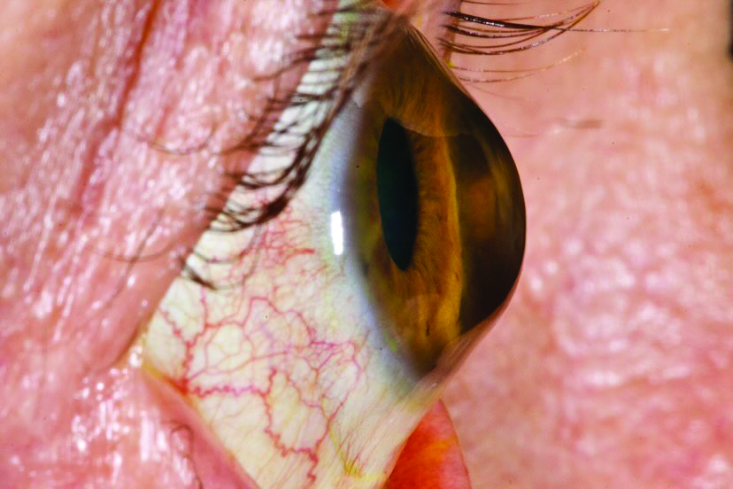
New research reveals that ocular effects of keratoconus even in early-stage disease are not exclusive to the anterior segment. Photo: Irving Martínez Navé. Click image to enlarge. |
While the effects of keratoconus on the anterior segment are often the focus of clinical studies, new research shines light on the potential impact of the disease on the back of the eye. Even in its early stage, researchers found evidence that keratoconus may cause bowing of the lamina cribrosa, subtle peripapillary retinal nerve fiber layer (RNFL) thinning and vascular alterations.
The single-center investigation evaluated 32 eyes with keratoconus (97% with early-stage disease) and 32 age- and sex-matched controls. The researchers performed various scans on the cohort including anterior segment OCT, Placido-disc topography, macular and optic nerve head swept-source OCT (SS-OCT), SS-OCT-A scans and a 3D wide glaucoma module for measuring peripapillary RNFL thickness.
Patients with keratoconus appeared to have a reduced peripapillary RNFL thickness compared with controls (104.8µm vs. 110.7µm), as well as reduced nerve radial peripapillary capillary plexus vessel density (46.3% vs. 43.8%). Especially in the temporal sector, these differences were more evident. These two observations were also associated with a higher lamina cribrosa curvature index in keratoconus patients vs. controls (9.9% vs. 8.5%). Another significant finding: in keratoconus eyes, macular superficial capillary plexus vessel density was 2.74% lower than controls.
The researchers offered a possible explanation for these findings in their paper on the study, recently published in American Journal of Ophthalmology. “Although no histological data are available, lamina cribrosa may be altered in keratoconus either due to its collagenous structure or through deformation of the whole posterior sclera and its peripapillary anchor to lamina cribrosa (scleral flange),” they wrote.
Although the central macular and choroidal thickness appeared to be unaffected by the presence of keratoconus, the researchers speculated that this may be due to the inclusion of only patients with early-stage disease in the study. It may also be a result of the software-based measurement technique used for calculation. Additionally, the study’s small sample size should also be taken into account when interpreting these findings.
The researchers concluded, “Early-stage keratoconus may be characterized by a posterior bowing of lamina cribrosa along with a subtle peripapillary RNFL thinning and vascular impairment.” Additionally, they pointed out that the study’s findings support the hypothesis that the disease could be a corneal manifestation of a more generalized eye collagen disease, “which would also affect collagenous structures of the posterior pole, such as peripapillary sclera, lamina cribrosa and retinal vessel wall.”
Pierro L, Bianco L, Bertuzzi F. New findings in early-stage keratoconus: Lamina cribrosa curvature, retinal nerve fiber layer thickness and vascular perfusion. Am J Ophthalmol. October 30, 2022. [Epub ahead of print]. |
