 |
History
A 76-year-old black male was referred to our group practice for cataract extraction due to decreased vision. The patient had a documented history of mild primary open angle glaucoma, which was being monitored by the referring doctor. The patient was known to be noncompliant with topical therapy and had a history of laser trabeculoplasty performed in both eyes twice before. The patient had a systemic medical history of hypertension, controlled with anti-hypertensive medications. He reported no allergies to medications or food.
Diagnostic Data
His best-corrected visual acuity measured 20/100 OD, 20/40 OS; interferometer acuity was 20/25 OU. External examination was unremarkable. Pupils were equal, round and reactive to light with a negative APD. Anterior segment evaluation revealed corneal arcus 360 degrees in both eyes, clear conjunctiva, flat and round iris and a deep and quiet anterior chamber. Upon evaluation of the lenses, the patient had grade two nuclear sclerosis in both eyes, along with cortical spoking greater in the right eye than in the left. Intraocular pressures were measured as 22mm Hg OD and 21mm Hg OS. Posterior segment findings revealed mild attenuation of vessels secondary to hypertension. Optic nerve head, macula, posterior pole and periphery were unremarkable. The patient was referred for a combination procedure and underwent phacoemulsification of the lens with IOL implant and the insertion of an iStent (Glaukos) in each eye.
Postoperative visits begin by checking for patient compliance regarding the postoperative regimen; we make sure the patient takes the prescribed topical drops, engages in no activities that involve bending or lifting and wears an eye shield or sunglasses, or both, outdoors. The examination usually includes a measurement of visual acuity, checking pupils, checking the anterior segments, measuring IOP and an undilated inspection of the posterior poles. Postoperative evaluations traditionally occur at one day, one week, three weeks and eight weeks.
Upon a second postoperative evaluation, approximately two weeks after the procedure, the patient’s IOP was measured to be 28mm Hg OD.
Your Diagnosis
Does this case require any additional tests? What is your diagnosis? How would you manage this patient? What’s the likely prognosis?
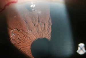 | |
| Gonioscopy image of a 76-year-old black male patient with glaucoma drainage implant. |
Discussion
The iStent trabecular micro-bypass is a titanium stent placed ab interno (Internal trabecular meshwork access), by passing the trabecular meshwork.1 This procedure is typically done in conjunction with cataract surgery as the stent can be placed into the trabecular meshwork via one of the same corneal incisions. With 75% of the outflow resistance found in the juxtacanicular tissue, a single iStent can improve outflow by 84%.2
Minimally invasive glaucoma surgeries (MIGS) are a novel alternative for patients who need cataract surgery and have a history of non-compliance with glaucoma medications. MIGS allows mild to moderate primary open-angle glaucoma patients better intraocular pressure (IOP) control with or without drops. While MIGS procedures are simple and require only modest skill, they do not decrease the IOP as dramatically as a trabeculectomy shunt (TVT).3 The advantage of a MIGS procedure is decreased complication rate compared with either a trabeculectomy or tube placement.3 Investigators report that the iStent has virtually no risk of bleb leak, bleb-related infections or hypotony. Currently, FDA-approved MIGS procedures include the Ex-Press Mini Glaucoma Shunt (Alcon) and the iStent (Glaukos).1-3
A study assessed the safety and efficacy of the iStent in combination with cataract surgery, in patients with mild and moderate primary open-angle glaucoma.3 It found that 60% of treated eyes had more than a 20% reduction compared with a rate of 48% of controls reaching that level of reduction.3 A potential source of bias is that cataract surgery alone independently helps to decrease IOP. Another study found that the success rate between the Ex-press filtration device (an ab externo procedure—placed from outside the trabecular meshwork) which is placed under a scleral flap through the trabecular meshwork into the Schlem’s canal without unroofing it, is comparable to a standard trabeculectomy.4 The advantage of this procedure is that fewer complications have been reported compared with standard trabeculectomy.
Investigators report IOP control is similar between the standard trabeculectomy and the Ex-press shunt, even five years after surgery.5
The gonioscopy view of the right eye demonstrates a displaced stent. The stent was still attached to the meshwork, but was not filtering the aqueous into the canal of Schlemm. Depending on the final position of the stent, the surgeon can either leave it or remove it altogether from the anterior chamber. Failure to implant the device in the trabecular meshwork can cause it to be displaced and touch the iris, contact the corneal endothelium or remain mal-positioned and poorly functioning. In these cases, it can either be repositioned, in an outpatient procedure or the iris can be contracted with a laser burn (iridoplasty).
Day one post-operative exams can reveal mild hemorrhaging at the placement site, resolving in about a week.
Despite minor complications occurring with minimally invasive glaucoma surgeries, they are gaining popularity among glaucoma and cataract surgeons. With less morbid complications and comparable results to tube shunts and a trabeculectomy, MIGS offer a reasonable first alternative to invasive procedures.
Dr. Gurwood thanks Kriti Bhagat, OD for contributing this case.
1. Nichamin L. Glaukos iStent Trabecular Micro-Bypass. Middle East African Journal of Ophthalmology 2009;16(3):138-140.2. Craven ER, Katz L, Wells JM, Giamporcaro JE. Cataract Surgery with trabecular micro-bypass stent implantation in patients with mild-to-moderate open-angle glaucoma and cataract: Two-year follow up. Journal of Cataract & Refractive Surgery 2012;38(8):1339-1345.
3. Sameulson TW, Katz LJ, Wells JM et al. Randomized Evaluation of the Trabecular Micro-Bypass Stent with Phacoemulsification in Patients with Glaucoma and Cataract. Ophthalmology 2011;118(3):459-467.
4. Maris PJ., Ishida K, Netland PA. Comparison of trabeculectomy with Ex-PRESS miniature glaucoma device implanted under scleral flap. Journal of Glaucoma 2007;18(1):14-19.
5. de Jong, LA. The Ex-PRESS glaucoma shunt versus trabeculectomy in open-angle glaucoma: a prospective randomized study. Advances in Therapy 2009; 26(1);336-345.
6. Samuelson TW. Microinvasive glaucoma surgery - Coming of age. Journal of Cataract & Refractive Surgery 2014;40(8):1253-1254.
