Q: I recently began comanaging glaucoma cases. What steps can I take to insure that I am protecting the patient and myself as I manage their disease over a period of years?

A: The short answer: Take all the steps. Keep on top of your patients and don’t cut corners. That means being timely with their visits and tests, as well as being meticulous with your documentation and records.
Let’s step back a moment: First, bear in mind that glaucoma is usually a very slowly progressing disease. There’s no need to hurry into a diagnosis or treatment decision.
But having said that, also know that it is a very serious disease, which means we have to take it seriously, every step of the way. You don’t want to overlook anything that might allow you to miss or delay the diagnosis.
With that in mind, I recommend you do what we do in our office: Have a “cheat sheet” of tests and procedures. In each glaucoma patient’s record, we have a handy one-page top sheet that allows the doctor to review the results of every visit and diagnostic test at once. It not only shows you the patient’s record at a glance, but it also serves as an easy reminder not to skip or postpone any important elements of the patient’s exam. Similarly, it encourages you to have a standard protocol of testing and evaluation for all your glaucoma patients.
We include these results on our flow sheet:
• Date of visit.
• Time seen.
• Time medication was last used.
• Visual acuity.
• IOP. A note about taking intraocular pressures: It’s important for the doctor to be the one who takes the pressures, or at the very least spot-checking them often. Taking accurate Goldmann IOP readings is not a “no brainer,” so do all you can to insure that they are accurate and consistent.• Visual fields. Like IOPs, the doctor should make sure that visual fields are performed properly. Make sure patients get encouragement and instruction. I’ve seen patients left in the visual field room for 10 minute stretches without anyone attending to them. To get the most reliable information from this very subjective (and very boring) test, keep your staff well trained and in and out of the field room to encourage patients as they go through it.
• Fundus appearance. Look at the disc in stereo, then take a photo. But even with a photo, carefully draw the disc. (Nothing wrong with taking disc photos, but drawing the disc encourages you to really think about the size, shape, rim tissue and cupping with every eye of every patient.) Also, while photos are helpful for your records, they are two-dimensional; they don’t adequately represent the excavation of the disc. You need to view that disc stereoscopically at appropriate intervals.
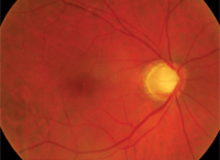
You can be fooled if you don’t spend enough time examining the optic nerve.
• Gonioscopy. This is a very critical part of the glaucoma evaluation, but is not done nearly as frequently as it should be. In order to diagnose open or closed angle glaucoma, one has to look into the angle and not just estimate the chamber depth.
• OCT/GDX. These instruments are a great addition to our glaucoma toolbox. But in order to provide top-notch glaucoma care, don’t let the results lull you into not looking at the optic nerve. Use all the information you have gathered from the exam to come up with your impression and plan.
In short, to ensure quality patient care, look at every patient’s nerve as if they have glaucoma until proven otherwise. Don’t rely on IOP. Don’t rely on a single test.
Don’t be lulled into complacency even if their IOP is stable. Track these patients to know if they are following up with their visits and compliant with your treatment. Be thorough, treat your patients well, and treat their records even better!
If you are doing the right things the right way each and every time, then you can rest assured that your patients are getting the care they need and deserve.
