Technicians should be permitted to perform subjective refractions.
By JAMES K. KIRCHNER, O.D.  Technicians should be permitted to perform subjective refractions for two reasons: Optometric scope of practice continues to evolve, and technology has enabled technicians to perform the data-gathering portion of the refraction effectively.
Refraction is both an art and a science. The mechanical steps that are required to perform a refraction are the science. These mechanical steps include using combinations of lenses to find the best endpoint that provides the patient with clear and relaxed vision. With the development of the phoropter, the science of knowing how to combine lenses to focus light on the retina was enhanced. We learned to correct astigmatism, hyperopia, myopia, presbyopia and muscle imbalances using a phoropter.
The interpretive and subsequent prescribing elements are the art. These include incorporating other factors, such as patient history gathered from the examination and interpreting and analyzing the data gleaned from the refraction, so that we can create a therapeutic plan of action, develop the prescription and clearly communicate the results to the patient. Both factors comprise a complete eye exam.
Some who are against delegating subjective refraction might assume that optometrys evolving scope of practice has overshadowed the professions beginnings, and that the quality of care diminishes when a technician oversees the mechanical aspect of the refraction. My contention, however, is that patients quality of care actually improves.
Scope of Practice
Refraction is a unique service that optometry has primarily provided and should continue to provide. Yet, as a profession, we learned to appropriately delegate frame selection, contact lens insertion and removal training, and more to trained staff. Why? So we could evolve from rudimentary evaluations and eyeglass sales into full-scope healthcare providers who deliver comprehensive eye care, including the diagnosis and treatment of various ocular diseases.
About five years ago, my partners and I decided that, if we wanted to grow our practice, we would have to become more efficient. We felt strongly that improved efficiency would increase the quality of care for our patients. We felt it would decrease waiting time for patients throughout the complete visit. We could delegate certain tasks to staff, who could take the time to perform the duty more thoroughly, allowing us to both concentrate on the patients needs by spending extra quality time with them and introduce more services than we previously had.
So, we looked at each step of the patient encounter. In analyzing how we spent our time in clinic, for instance, we realized that a lot of professional time was spent in the act of the refraction. And, because we were spending so much time on the science aspect, we were not spending enough time talking to patients about their results. There- fore, one of the steps we took to streamline our practice was to delegate the science part of the refraction to our technicians.
Technology
While some O.D.s feel a staff person does not have the educational background to perform the mechanical portion of a subjective refraction, I believe that computer-assisted subjective refractions have made this argument obsolete.
Computer-assisted subjective refraction is a completely different item compared to automated objective refractions. Our personal experience has shown that this equip- ment allows a technician to perform a refraction that equals or possibly exceeds a standard phoropter refraction.
In our practice, we use the Marco Epic 2100 system. The system takes a trained technician through a step-by-step sequence that is programmed and consistent. In combination with an autolensometer and an autorefractor, data is delivered to the Epic phoroptor. The phoropter is a full-scale unit, but its electronically driven so that it is easier to control. A technician then has all the tools to do a complete, accurate subjective refraction.
Each step of the refractive sequence is pre-programmed, and you cannot move to the next step without accomplishing the one you are at. First, acuities are taken aided or unaided, then the phoropter loads the autorefractive data. At this point, the refraction routine begins. Correction for sphere, cylinder and axis is determined for each eye. If chosen, a binocular refraction with over-plus on the non-tested eye can be done. After the correction is determined, phorias or a fixation disparity can be tested. All the lenses and testing targets are available.
Finally, an instant comparison between the habitual Rx and the new one can be done to demonstrate the additional clarity of the new Rx to the patient. This combination testing with the computer programming results in a very accurate outcome.
Once this refractive sequence is completed, I have the new results to compare with the previous Rx, the auto refraction data, the patients complaints and the patients history all at my disposal. Then, I can accept or modify the Rx using my best clinical experience.
In a few cases, when one piece of data doesnt correlate with another, I recheck it using the standard phoropter. For instance, there could be a case in which the new cylinder axis doesnt appear to be in harmony with the old Rx or the auto refraction. Or, a young hyperope may have less plus on one eye compared to what you would have assumed should be present. But these instances occur less than 15% of the time.
My system also allows the patient to see the targets simultaneously, enabling a very accurate comparison and choice. This aspect has proven to be a huge benefit in refracting patients who have lower acuities as a result of macular degeneration or cataracts.
There have been times in which weve had problems getting patients who have some physical handicaps into the instrument. When this happens, we opt for the traditional mode of refracting.
My partners and I were initially concerned that patients would react negatively to our technicians conducting the scientific aspect of the refraction, but the opposite has occurred. Some patients have said they felt they were getting better service and were impressed with the technology. They are also happy that we were now able to spend more quality time with them to discuss their needs, concerns and options.
In the end, from our long-term experience, we know that a technician can produce the same end results as we would using traditional methods. An added benefit is a boost in staff morale. Our technicians are excited to learn this new realm of testing.
The final analysis and decision- making should be done by the O.D., because only we know how that data will benefit our patients. But, the data-gathering portion can be delegated. And, I think it is egotistical for us to believe that we are the only people who can learn to do a refraction. Skilled, trained technicians are eager and able to learn this procedure.
Optometry has a great future, but we must know what to hold on to and what we can delegate so that we have the time to provide all the services that will be available to our patients in the future. n
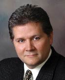
Dr. Kirchner is the founder and senior partner of a group, multi-office practice in Lincoln, Neb. He predominately concerns himself with primary-care optometry, with an emphasis on contact lenses and the treatment of eye diseases.
Only the optometrist should be permitted to perform subjective refraction.
By MERRILL D. BOWAN, O.D.  Opticians and technicians should not be permitted to perform subjective refractionswith or without automated technologyfor two reasons: They do not have the necessary educational background to best serve patients, and without that background, the information they obtain is meaningless and can even be detrimental to patients welfare.
Some who favor opticians refracting might assume that all an O.D. needs to satisfy a patients visual requirements are the right numbers in the LCD display, in the phoropter windows or in the prin- ter output. However, not all refractions are the same.
Other Factors
Indeed, several factors can influence the outcome of the refraction, including:
- Stress. Stress can manifest itself in the refraction as a result of the emotional distraction of the patient or from cortisolic effects on the index of refraction of the lens.1-3
For example, years ago, I had a female patient come in for a routine refraction, and her responses were all over the dial. She varied from
-0.50D to -0.75D in cylindrical power, the axes swung across a 20-degree range, her hyperopic correction could not be pinned down within a 0.50D range, and her visual acuities never got better than 20/30+/-. I recommended that she come back in a week, and the next visit went like clockwork. The patient manifested moderate hyperopic astigmatism. When she asked why there was such a difference, I suggested that stress may have played a role. Sure enough, she divulged that at the time of the last visit, her dog was going in for surgery the next day. - Medical conditions. Conditions such as hyperthyroidism and blood sugar variations can alter a refraction.4-9 Hyperthyroidism is also known to alter extraocular muscle functions, affecting convergence, vertical imbalances and the corneas integrity.9
Refractive changes are often the first indication of diabetes mellitus, particularly of the growth-onset variety.9 There is one personal case of diabetes mellitus that comes to mind. Prior to the same-day-glasses era, a patient who suffered a hyperglycemic crisis came directly from the hospital to my office. His 5.00D lens prescription was too strong by three diopters. I advised him to wait three weeks for his insulin levels to balance out. When he told me he could not wait and had to work, I acquired a pair of 3.00D readers, removed the lenses and secured them to the front of his habitual lenses. I saw him a week later, and his refraction had mostly recovered to the previous level. At that point, I wrote him a new prescription, though I cautioned that the Rx might change further. Nevertheless, he was willing to risk the changes. Two weeks later, we checked the refraction once more, and it had barely changed. - Neurodevelopmental issues. Given that my specialty is in neurodevelopmental visual issues, I see many children who have school-related vision problems. These vision problems affect the whole behavioral spectrum, resulting in visual processing-based problems in learning disabilities, ADD/ADHD and even autism.10-16 Studies have also shown that low to moderate hyperopia correlates with visual-motor problems.17-19 In addition, patients with learning disabilities often refract at near-emmetropic ranges, are shown to have high levels of neural hyper-reactivity and can benefit from ophthalmic lenses and prisms.20-23 These types of problems will not present in a data-based-only exam. Only the O.D. is aware of the academic abilities, reading styles, aversions to nearpoint work (including symptomatology) and handwriting skills of these children, all of which affect the Rx.
The bottom line is that only the educated O.D. understands that a visual exam can be affected by stress, medical conditions, neurodevelopmental issues and more. And the O.D. (not the optician or technician) understands how these patients measurements relate to their performance, responses and needs. Vision dominates the other senses. If we fail to understand this, we risk short-changing the quality of our patients lives.
Recently, after treating an 11-year-old girl who has a vision-related learning disability, her mother said, My daughters changes are not just academic, she actually seems calmer, more secure. These changes in quality-of-life will never be accomplished by a technician, even with a subjective autorefractor. The best clinical techniques optometry has to offer changed this childs vision and, therefore, her view of the world.
Scope of Practice
We cannot lose sight of what defines a patients needs and why an eye examination is necessary. While we have a medicolegal responsibility to identify, refer or treat ocular medical conditions, we cannot ignore our accountability to identify, refer or treat the 20% to 32% of the population who manifest binocular or developmental dysfunctions.24-26
Refraction and visual analysis are road tests of the visual system under controlled conditions. Does the patients visual system have the resources to cope, as measured by the duction tests? Is it distorting due to performance stresses, as reflected by the phorias? Has it begun to collapse, as in Streff Syndrome?
Optometry is the only profession capable of bridging visual functioning and visual perceptual processing with the manifestation of any external distortions. We cannot ignore what comprises a true refraction. This issue must be resolved within optometry not so much for our profession, but for the publics welfare.
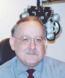
Dr. Bowan is a private optometric clinician with a specialization in neurodevelopmental visual issues. He practices in the Greater Pittsburgh area.
- Asvetisov ES, Gundorova RA, Shakarian AA, et al. [Effects of acute psychogenic stress on the state of several functions of the visual analyzer.] Vestn. Oftalmol. 1991 Jan-Feb, 107(1):17.9.
- Weinstein P, Dobossy M. The psychosomatic factors in ophthalmology. Klin Monatsbl Augenheilkd. 1975 Apr;166(4):537-9.
- Gawron VJ. Ocular accommodation, personality, and autonomic balance. Am J Optom Physiol Opt. 1983 Jul; 60(7):630-9.
- Kinoshita S. [Pathogenesis and treatment of accommodative disturbance.] Nippon Ganka Gakkai Zasshi. 1994 Dec;98(12):1256-68.
- Huismans H. [Conspicuous change of refraction in endocrine orbitopathy.] Klin Monatsbl Augenheilkd. 1991 Mar;198(3):215-6.
- Fledelius HC. Refractive changes in diabetes mellitus around onset or when poorly controlled. A clinical study. Acta Ophtalmol (Copenh). 1987 Feb; 65(1):53-7.
- Eva PR, Pascoe PT, Vaughan DG. Refractive change in hyperglycaemia: hyperopia, not myopia. Br J Ophthalmol. 1982 Aug;66(8):500-5.
- Giusti C. Transient hyperopic refractive error changes in newly diagnosed juvenile diabetes. Swiss Medical Wkly. 2003 Apr 5;133(13-14):200-5.
- Newell FW. Ophthalmology. Principles and concepts. Fourth Edition. CV Mosby. St. Louis. 1978. (Chapter 25.)
- Evans BJ, Drasdo N, Richards IL. An investigation of some sensory and refractive visual factors in dyslexia. Vision Res. 1994 Jul;34(14):1913-26.
- Buzzelli AR. Stereopsis, accommodative and vergence facility: do they relate to dyslexia? Optom Vis Sci. 1991 Nov;68(11):842-6.
- Maples WC, Bither M. Efficacy of vision therapy as assessed by the COVD quality of life checklist. Optometry. 2002 Aug;73(8):492-8.
- Raggio DJ. Visuomotor perception in children with attention deficit hyperactivity disorder-combined type. Percept Mot Skills. 1999 Apr;88(2):448-50.
- Kaplan M, Edelson SM, Seip JA. Behavioral changes in autistic individuals as a result of wearing ambient transitional prism lenses. Child Psychiatry Hum Dev. 1998 Fall; 29(1):65-76
- Carmody DP, Kaplan M, Gaydos AM. Spatial orientation adjustments in children with autism in Hong Kong. Child Psychiatry Hum Dev. 2001 Spring; 31(3):233-47.
- Denis D, Burillon C, Livet MO, et al. [Ophthalmologic signs in children with autism.] J Fr Ophthalmol. 1997;20(2):103-10.
- Rosner J, Rosner J. The relationship between moderate hyperopia and academic achievement: how much plus is enough? J Am Optom Assoc. 1997 Oct;68(10):648-50.
- Rosner J, Gruber J. Differences in the perceptual skills development of young myopes and hyperopes. Am J Optom Physiol Opt. 1985 Aug;62(8):501-4.
- Rosner J, Gruber J. Differences in the perceptual skills development of young myopes and hyperopes. Am J Optom Physiol Opt. 1985 Aug;62(8):501-4.
- Bowan MD. Introducing the binocular dissonance test. Presentation at the 65th Middle Atlantic Congress. Pittsburgh. September 20-21, 2003.
- Evans JR. Relationships between visual skills and behavior disorders. J Am Optom Assoc. 1968 Jul;39(7):632-40.
- Turatto M, Facoetti A, Serra G, et al. Visuospatial attention in myopia. Brain Res Cog Brain Res. 1999 Oct 25;8(3):369-72.
- Sucher DF, Stewart J. Vertical fixation disparity in learning disabled. Optom Vis Sci. 1993 Dec;70(12):1038-43.
- Porcar E, Martinez-Palomera A. Prevalence of general binocular dysfunctions in a population of university students. Optom Vis Sci. 1997 Feb; 74(2):111-3.
- Lara F, Cacho P, Garcia A, et al. General binocular disorders: prevalence in a clinic population. Ophthalmic Physiol Opt. 2001 Jan;21(1):70-4.
- Hokoda SC. General binocular dysfunctions in an urban optometry clinic. J Am Optom Assoc. 1985 Jul;56(7):560-2.

