“The death of nerve cells is the key event in all neurodegenerative disorders but, until now, it has not been possible to study cell death in real time,” says lead author M. Francesca Cordeiro, M.D., Ph.D. “This technique means we should be able to directly observe retinal nerve cell death in patients, which has a number of advantages in terms of effective diagnosis. This could be critically important since identification of the early stages could lead to successful reversal of the disease progression with treatment.”
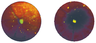
The retina of an Alzheimer's transgenic mouse (left) shows many more retinal nerve cells in the early phase of apoptosis (green spots) vs. the retina of a control mouse (right).
The technique uses fluorescent markers that attach to the relevant cells and indicate the stage of cell death. The retina is then observed using a customized confocal scanning laser ophthalmoscope. This permits the tracking of changes in the same cell in the same eye over hours, days, weeks and months in acute, chronic and treated glaucoma and Alzheimer models of neurodegeneration.
This research is the first ever in vivo demonstration of retinal nerve cell death in Alzheimer’s disease.
Because neurodegenerative diseases, such as Alzheimer’s and glaucoma, have mechanisms in common, tracking the dynamics of cell death in one disease could enhance the understanding of other diseases, in terms of both the molecular pathophysiology and the potential therapeutic avenues, the researchers write.
“The equipment used for this research was customized to suit animal models, but is essentially the same as is used in hospitals and clinics worldwide,” Dr. Cordeiro says. “It is also inexpensive and non-invasive, which makes us fairly confident that we can progress quickly to its use in patients.”
To that end, the researchers will be conducting their first patient trials later this year.
“Few people realize that the retina is a direct, albeit thin, extension of the brain,” Dr. Cordeiro says. “It is entirely possible that, in the future, a visit to a [neighborhood optometrist] to check on your eyesight will also be a check on the state of your brain.”
For more, see this month's Comanagement Q+A: “
Keep Alzheimer’s in Mind."
