A 35-year-old black female presented for a routine eye examination. She had no ocular complaints, signs or symptoms. Her systemic history was unremarkable. She reported using no medications and has no known allergies. Diagnostic Data Her best-corrected visual acuity was 20/20 O.U. at distance and near. External examination was normal, with no evidence of afferent pupillary defect. Refraction revealed myopia consistent with her current spectacle correction. Intraocular pressure measured 19mm Hg O.U. How would you approach this case? Does this patient require any additional tests? What is your diagnosis? How would you manage this patient? Whats the likely prognosis? Discussion The diagnosis in this case is arteriovenous (AV) malformation. Additional tests might include fundus photography to record the appearance and size of the anomaly, laser interferometry to assess the maximum potential for foveal acuity if vision is reduced, and visual field testing to rule out visual deficits. Neuroimaging or neurosurgical referral must be considered because central nervous system arteriovenous malformations also may exist. 1. Gurwood AS, Bailey JT, Pelino CJ. Congenital retinal macrovessel: a case report. Optometry 2001 Sep;72(9):597-602. 2. Meinhold JR. Arteriovenous communications of the retina. J Am Optom Assoc 1996 May;67(5):279-82. 3. Beatty S, Goodall K, Radford R, Lavin MJ. Decompensation of a congenital retinal macrovessel with arteriovenous communications induced by repetitive rollercoaster rides. Am J Ophthalmol 2000 Oct;130(4):527-8. History
The pertinent posterior segment findings are demonstrated in the photograph.
Your Diagnosis
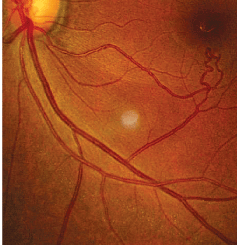
The right eye of our 35-year-old patient who presented for a routine examination. Though she voiced no ocular complaints, do you notice anything suspicious?
Arteriovenous communication of the retina (AVCR) is a rare, congenital retinal vascular anomaly.1-3 It is usually present unilaterally, with a predilection for the papillomacular area and superotemporal quadrant of the retina.2 Men and women are equally affected by AVCR, and the condition is rarely inhereted.1-3 Visual acuity is usually unaffected, and associated systemic AV communications are atypical in grade I or II AVCR.2 Grade III AVCR is the most severe variant of the condition, and it is often linked with mid-brain AV malformations.2
Congenital retinal macrovessels are rare, aberrantly large branches of retinal arteries or veins that typically cross the horizontal raphe and either supply or drain the macular area.1 These unusual vascular entities must be distinguished from prepapillary vascular loops, racemose hemangiomas, Wyburn-Mason syndrome, or other arteriovenous malformations that have potential neuro-ocular sequelae.1
Though most of these vascular anomalies remain intact with an uncomplicated course throughout the patients life, certain activities that exert sudden or repetitive g-forces, such as bungee jumping and rollercoaster riding, may pose an increased risk of retinal vascular decompensation.3
We educated our patient about the condition and evaluated her for additional complications using neuroimaging. To date, she has experienced no deleterious complications or changes.
