 History
A 47-year-old black female presented for a routine eye examination. She reported no visual or ocular complaints. Her systemic history was noncontributory. She took no medications and reported no allergies of any kind.
Diagnostic Data
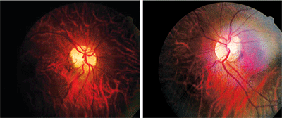
Her best-corrected visual acuity measured 20/20 O.U. at distance and near. External examination was normal, with no evidence of afferent pupillary defect. Her anterior segment findings were normal. Intraocular pressure was 18mm Hg O.U. The pertinent fundus findings are illustrated in the photographs.
 |
| This 47-year-old woman presented for a routine eye exam. Do you notice any significant fundus findings (O.D. left, O.S. right)? |
Your Diagnosis
How would you approach this case? Does this patient require any additional tests? What is your diagnosis? How would you manage this patient? What’s the likely prognosis?
Diagnosis
The diagnosis in this case is anomolous discs. Additional testing might include Amsler grid, central threshold visual fields to rule out deficits, photodocumentation, and monitoring for the development of peripapillary subretinal neovascularization.1 Ophthalmoscopically, many patients exhibit considerable variation in optic disc size. The size and shape of an optic disc can be influenced in part by refractive error. At times, it can be difficult to determine if the size of the disc falls inside or outside the normal limits.
Megalopapilla, an abnormally large optic disc, is exceedingly rare.2,3 Micropapilla is more common, however, and warrants inclusion in the differential diagnosis of any child with poor vision in one or both eyes. Other congenital disc malformations are often accompanied by field defects that may be mistaken for an acquired disease. Or, the defects can be regularly misinterpreted as nerve head swelling. Occasionally, this causes patients to undergo unnecessary diagnostic procedures, such as magnetic resonance imaging.4
Differential diagnoses of an anomalous disc appearance include posterior staphyloma (scleral outpouching), coloboma (congenital notch), morning glory disc (congenital fibroglial abnormality), optic disc drusen and hypoplastic disc (small disc secondary to decreased axons).1-10 We educated this patient regarding the uniqueness of her presentation and our rationale for capturing photo-documentation. Because there was no evidence of fluid leakage, choroidal neovascularization or an optic pit, direct intervention was unnessessary.1-8
1. Alexander LT. Congenital and acquired anomalies of the optic nerve head. In: Alexander LS. Primary Care of The Posterior Segment. Appleton & Lange: Norwalk, Conn., 1994:89-170.
2. Swann PG, Coetzee J. Megalopapilla. Clin Exp Optom. 1999 Sep-Oct;82(5):200-2.
3. Sampaolesi R, Sampaolesi JR. Large optic nerve heads: megalopapilla or megalodiscs. Int Ophthalmol. 2001;23(4-6):251-7.
4. Sowka JW, Luong VV. Bitemporal visual field defects mimicking chiasmal compression in eyes with tilted disc syndrome. Optometry. 2009 May;80(5):232-42.
5. Lempert P. Optic nerve hypoplasia and small eyes in presumed amblyopia. J AAPOS. 2000 Oct;4(5):258-66.
6. Berk AT, Yaman A, Saatci AO. Ocular and systemic findings associated with optic disc colobomas. J Pediatr Ophthalmol Strabismus. 2003 Sep-Oct;40(5):272-8.
7. Beuchat L, Safran AB. Optic nerve hypoplasia: papillary diameter and clinical correlation. J Clin Neuroophthalmol. 1985 Dec;5(4):249-53.
8. Chan RT, Chan HH, Collin HB. Morning glory syndrome. Clin Exp Optom. 2002 Nov;85(6):383-8.
9. Doyle E, Trivedi D, Good P, et al. High-resolution optical coherence tomography demonstration of membranes spanning optic disc pits and colobomas. Br J Ophthalmol. 2009 Mar;93(3):360-5.
10. Spencer TS, Katz BJ, Weber SW, et al. Progression from anomalous optic discs to visible optic disc drusen. J Neuroophthalmol. 2004 Dec;24(4):297-8.

