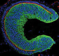
Human embryonic stem cells (hESC) can be used to grow tissue in the form of a complete optic cup, according to a new study in the June 14 edition of Cell Stem Cell. In the future, transplantation of this 3D tissue could restore sight to visually impaired patients.

A human embryonic stem cell-derived optic cup
generated in culture. Bright green is neural retina;
off green is pigment epithelium; blue is nuclei; and
red is active myosin.
“Our approach opens a new avenue to the use of human stem cell-derived complex tissues for therapy, as well as for other medical studies related to pathogenesis and drug discovery,” says senior study author Yoshiki Sasai of the RIKEN Center for Developmental Biology in Japan.
The researchers optimized cell culture methods that enabled the hESC-derived cells to form the correct 3D shape and the two layers of the optic cup, including a layer of photoreceptors. Because retinal degeneration typically results from damage to these cells, this tissue could be ideal for transplantation in the future.
The same research group previously created an optic cup derived from mouse embryonic stem cells. But the hESC-derived optic cup seems to contain species-specific instructions for building an ocular structure. It’s much larger in size and its multilayered tissue contains both rods and cones, which is rare in mouse ESC cultures.
Nakano T, Ando S, Takata N, et al. Self-formation of optic cups and storable stratified neural retina from human ESCs. Cell Stem Cell. 2012 June 14;10(6):771-85.

