 History
A 37-year-old black male presented for an eye examination following hospitalization for an advanced HIV/AIDS infection. The internist referred the patient to us to rule out significant retinopathy.
The patient presented with a chief complaint of decreased visual acuity in his left eye. Additionally, he reported irritation and photophobia in both eyes that had persisted for three days.
The patient explained that he had been rubbing his eyes aggressively. He denied any relevant ocular history and was being medicated for HIV with ganciclovir. He reported no allergies.
Diagnostic Data
Best-corrected visual acuity measured 20/30 O.D. and 20/200 O.S. at distance and near. External examination was normal, with no evidence of afferent pupillary defect. The palpebral and bulbar conjunctiva were quiet and pink, without injection. There were no cells or flare in the anterior chamber. The pertinent anterior segment findings are illustrated in the photographs.
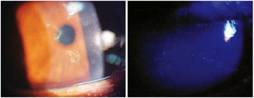 |
| This 37-year-old patient with advanced HIV presented with a chief complaint of decreased visual acuity in his left eye. What do you observe in the photographs?
|
Intraocular pressure measured 15mm Hg O.U. Fundus examination revealed HIV retinopathy (scattered dot hemorrhages with a few isolated cotton-wool infarcts), but no evidence of cytomegalovirus (CMV) retinitis, candidiasis or pneumocystis retinal infection.
Your Diagnosis
How would you approach this case? Does this patient require any additional tests? What is your diagnosis? How would you manage this patient? What’s the likely prognosis?
Additional testing included palpation of the nodes, checking the palpebral conjunctiva for follicles and papillae, eversion of the upper eyelids to rule out the presence of foreign matter, corneal sensitivity testing, tear evaluation for volume and quality (e.g., lacrimal lake observance, Schirmer test strips), and staining with sodium fluorescein and rose bengal. All tests were negative.
The diagnosis in this case is unclear and may never be determined. In fact, there was controversy and debate in the office among the group of practitioners.1-3 One camp believed that the presentation was simply an atypical corneal erosion/abrasion and recommended debridement, mild cycloplegia b.i.d., topical antibiotics q.i.d. and lubricants q.i.d. Given the patient's systemic history, the second camp believed the presentation reflected an atypical herpetic simplex infection and recommended mild cycloplegia b.i.d., topical Viroptic (trifluridine, GlaxoSmithKline) q2h and lubrication q.i.d.
After failing to reach a consensus, we contacted a corneal specialist and e-mailed the images seen above. But, the specialist was unable to identify the presentation and could not offer a definitive management plan. Ultimately, the second camp convinced the first camp of its diagnosis, and requested a follow-up with the patient in three days. No culture was obtained.
At follow-up three days later, we noted marked improvement. The patient reported feeling much better. He had almost no irritation and only mild photophobia. His best-corrected visual acuity had improved to 20/40 O.S.
However, upon checking the orders, I realized that the patient was not receiving the treatment we had prescribed. Instead, he received artificial tears q2h and Polysporin ophthalmic ointment (bacitracin zinc/polymyxin B sulfate, Monarch Pharmaceuticals) h.s. O.U. The attending physician explained that Viroptic was not on the formulary and had to be ordered.
The question remained: Was this patient suffering from an unusual corneal abrasion that was resolving secondary to conservative but appropriate management, or was he manifesting an atypical herpes simplex virus keratopathy that was resolving secondary to intravenous ganciclovir.
Despite the progress, we insisted on moving forward with the intended treatment (mild cycloplegia b.i.d., topical Viroptic q2h and lubrication q.i.d.). We also maintained the Polysporin ointment h.s. O.U. Again, we scheduled a follow-up examination three days later.
At follow-up, the lesion was 100% resolved. The acuity in his left eye had normalized to 20/25 and all the symptoms had faded. Did the Viroptic and ganciclovir combine to eradicate an atypical presentation of herpes simplex, or did the conservative treatment heal what was just a mild, superficial, mechanical corneal disruption? Without the culture, we will never know.
The patient will now remain on this “prescribed cocktail” to preserve his ocular and systemic health. Fortunately, the combination of medicines will also protect him against future outbreaks of herpes simplex virus.
1. Seitzman GD, Strauss EC, Margolis TP. "Steel wool keratopathy": a clinical sign of chronic inflammation.
Cornea. 2006 Jul;25(6):742-4.
2. Pilon AF, Scheiffle J. Ulcerative keratitis associated with crack-cocaine abuse.
Cont Lens Anterior Eye. 2006 Dec;29(5):263-7.
3. Garweg JG, Russ CE, Schellhorn M, et al. HSV-1 antigens and DNA in the corneal explant buttons of patients with non-herpetic or clinically atypical herpetic stromal keratitis.
Graefes Arch Clin Exp Ophthalmol. 2003 Sep;241(9):734-9.
