 The eye is continually exposed to organisms and other foreign molecules. To fight off these invaders, the bodys immune system has a whole army equipped with various defenses. The immune system is a coordinated network of cells, humoral factors, lymphoid organs and cytokines.
In this review, the first of two parts, we cover the normal function of the immune system in recognizing, repelling and eradicating pathogens.
Specialized Cell Types
The immune system is composed of many interactive cells that collectively protect the body from bacterial, parasitic, fungal and viral infections, as well as from the growth of tumor cells.1 The various cell types have specialized functions.
Lymphocytes. Lymphocytes are a family of cells characterized by a lack of specific granules. This cell type includes two major classes, the T and B lymphocytes.1,2 T lymphocytes (also known as thymus-dependent) are further divided into two major subsets: T-helper cells and
T-killer/suppressor cells.
T-helper cells (also called CD4+ cells) coordinate immune regulation by secreting specialized factors that activate other white blood cells to ward off infection.1 T-killer/suppressor cells (also called CD8+ cells) are involved in the direct killing of certain tumor cells, viral-infected cells and parasites. They are also important in the down-regulation of the immune response.1
Lymphocytes circulate with blood and lymph, and can infiltrate connective and epithelial tissues.2 To become stimulated, T lymphocytes must interact with histocompatibility molecules (membrane glycoproteins) that are present on the surface of eukaryotic cells and are coded by a set of genes known as major histocompatibility complex (MHC). Histocompatibility molecules belong to two types. The first is expressed on the surface of all cells. The other is exclusively expressed on the cells of the immune system.2
B lymphocytes (also known as bursa-dependent lymphocytes) are involved in the production of antibodies that respond to bacteria, viruses and tumor cells. Antibodies, also known as immunoglobulins, are specialized proteins that bind to a foreign substance (antigen) and signal other cells to engulf, kill or remove that substance from the body.1,2 Immunoglobulins are subdivided into several classes. The immunoglobins G (IgG) represent the bulk of the immunoglobulins in human blood. Other types include IgM, IgA, IgE and IgD.1,2
Natural killer (NK) cells. NK cells are similar to the T-killer cell subset (CD8+ T cells). They directly kill certain tumor (melanomas, lymphomas) and viral-infected cells (herpes and cytomegalovirus).1
Granulocytes. Granulocytes, or polymorphonuclear leukocytes (PMNs), are composed of three cell types: neutrophils, eosinophils and basophils, based on their staining characteristics. These cells remove bacteria and parasites from the body by engulfing and degrading them.1
Macrophages. Macrophages are often referred to as scavengers or antigen-presenting cells (APC) because they pick up and ingest foreign materials and present these antigens to other cells of the immune system (such as T cells and B cells). If stimulated, macrophages exhibit increased levels of phagocytosis and secretion.1
Dendritic cells. Dendritic cells originate in the bone marrow and function as antigen-presenting cells (APCs). They are more efficient APCs than macrophages and are usually found in lymphoid organs, such as the thymus, lymph nodes and spleen, as well as in the bloodstream.1
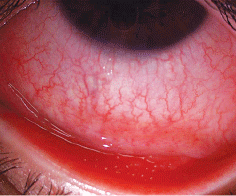 |
| Follicular conjunctivitis in adenoviral infection. |
Organs of the Immune System
The organs of the immune system include the bone marrow, thymus, spleen, tonsils and lymph nodes.
Bone marrow. The bone marrow produces all of the cells of the immune system, in addition to red blood cells and platelets, through a process called hematopoiesis. Bone marrow-derived cells either differentiate into mature cells (B cells) or precursors of cells that migrate elsewhere to continue their maturation (thymocyte/T cells).
Thymus. The thymus is responsible for producing mature T cells. Once the T cell is mature, it is released into the bloodstream.
Spleen. The spleen contains B cells, T cells, macrophages, dendritic cells, natural killer cells and red blood cells, and it is an immunologic filter of the blood. As blood passes through the spleen, antigens are captured. Macrophages and dendritic cells also present antigens to B and T cells in the spleen, initiating the immune response. In the spleen, B cells become activated and produce large amounts of antibody. Old red blood cells are destroyed.
Tonsils. Tonsils and adenoids are part of the lymphatic system. Tonsils are accumulations of diffuse, nodular lymphatic tissue found in the wall of the naso- and oropharynx. They help protect against infection by trapping germs coming in through the mouth and nose. Tonsils produce B cells and T cells.
Lymph nodes. The lymph nodes filter the bodily fluid known as lymph. Lymph nodes are composed mostly of T cells, B cells, dendritic cells and macrophages. The nodes drain fluid from most body tissues. Similar to the spleen, macrophages and dendritic cells capture antigens and present them to T and B cells, again initiating the immune response.1
Initiation of the Immune Response
The bodys immune response begins with an antigen-presenting cell (APC), usually either a macrophage or dendritic cell, presenting an antigen on its surface to a B cell.3 The B cell then proliferates and produces antibodies that specifically bind to that antigen. If the antibodies end up binding to antigens on bacteria or parasites, a signal is sent for PMNs or macrophages to engulf and kill them.
Antibodies also initiate the complement destruction cascade.1,3 Serum proteins, called complement, bind to immobilized antibodies and destroy the bacteria.3 Antibodies can also signal natural killer cells and macrophages to kill viral or bacterial-infected cells.
If the APC presents the antigen to T cells, the T cells become activated and proliferate. CD4+ T cells become secretory, and CD8+ T cells become activated to kill target cells that specifically express the antigen presented by the APC.1-3 The CD4+ T-helper cells regulate the production of antibodies and the activity of CD8+ T-killer cells. The CD4+ T cells provide growth factorsinterleukins and cytokinesto these cells and signal them to proliferate and function more efficiently.4 Interleukins and cytokines are crucial to ensure the activation of natural killer cells, macrophages, CD8+ T cells and PMNs.
Finally, both T and B lymphocytes have immunologic specificity. They are genetically programmed to respond to a specific antigen.2,4
Antigen Disposal
Two different mechanisms are responsible for antigen removal.
The first is the humoral immunological response.1 This mechanism, usually evoked by a bacterial antigen, allows lymphocytes to synthesize and release antibodies that attack the antigen.
The other mechanism is known as the cellular or cell-mediated immunological response (e.g., triggered by transplantation of foreign tissues). Cellular response causes clones of lymphocytes to either release molecules with pharmacological actions on macrophages, granulocytes or other lymphocytes, or attack the foreign cells directly.1,2
How does the immune response occur in the eye? Corneal infiltrates provide an example. Corneal infiltrates are a complication associated with contact lens wear. Infiltrates represent a cellular response resulting from tissue insult and inflammation. There is a migration of inflammatory cells, including polymorphonuclear leukocytes and mononuclear cells, from the limbal vasculature and precorneal tear film into the cornea, forming the infiltrate.
Next column (September): How immunomodulatory pharmacotherapies are used in clinical optometric practice.
1. Parkin J, Cohen B. An overview of the immune system. Lancet 2001 Jun 2;357(9270):1777-89. Review.
2. Raviola E. The Immune System. In: Fawcett DW, ed. Bloom and Fawcett: A Textbook of Histology. 12th ed.
3. Roitt I, Brostoff J, Male D. Immunology. 2nd edition.
4. Mosmann TR, Coffman RL. TH1 and TH2 cells: different patterns of lymphokine secretion lead to different functional properties. Annu Rev Immunol 1989;7:145-73.
