 A 79-year-old black male presented with a chief complaint of gradually decreased vision in his right eye, which started five months earlier. The eye had recently become mildly painful. He also reported a recent episode of dizziness and right-sided headache for which he sought treatment at a local emergency room. An MRI of the brain revealed no pathology.
His last eye examination was four years prior. Systemic history was remarkable for diabetes mellitus, hypertension, hypercholesterolemia and neuropathy in the lower extremities. The patient reported smoking five cigarettes a day. He was moderately obese. His most recent hemoglobin A1C was 8%. Systemic medications included 40mg Zestril (lisinopril, AstraZeneca), Glucotrol 10mg (glipizide, Pfizer), Plavix (clopidogrel, Bristol-Myers Squibb/Sanofi Pharmaceuticals) and TriCor 145mg (fenofibrate, Abbott). The patient was allergic to sulfa medications.
Best-corrected distance acuity was 20/50 O.D. and 20/25 O.S. Pupils were equally reactive with no afferent defect. Biomicroscopy revealed occasional anterior chamber cells and iris neovascularization without angle closure in his right eye, as well as bilateral lens opacities, worse in the right eye. Intraocular pressure measured 11mm Hg O.D. and 18mm Hg O.S. at 9:00 a.m. Dilated funduscopy revealed mid-peripheral dot-and-blot hemorrhages and microaneurysms, which were much more significant in the right eye.
Retinal veins were dilated but not tortuous. Retinal neovascularization was not seen on funduscopy or fluorescein angiography (FA), although FA of the right eye did show delayed filling in the arterial phase and areas of capillary non-perfusion. Optic nerves were healthy, with a cup-to-disc ratio of 0.50 x 0.50 in both eyes.
Blood pressure measured 140/92mm Hg, right arm sitting. Carotid auscultation demonstrated a “whooshing” sound consistent with bruit on the right side. We established a tentative diagnosis of hypoperfusion retinopathy (ocular ischemic syndrome). We initiated a topical steroid and a cycloplegic agent in the right eye to reduce inflammation and decrease the likelihood of hyphema.
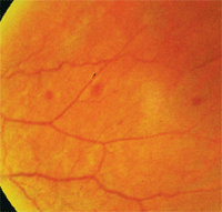
Mid-peripheral retinal hemorrhages are common in ocular ischemic syndrome.
We ordered carotid duplex-Doppler ultrasound and trans-thoracic echocardiogram, as well as erythrocyte sedimentation rate (ESR) and C-reactive protein (CRP). The carotid workup revealed stenosis of bilateral internal carotid arteries (95% right, 40% left). The patient soon underwent both panretinal photocoagulation in the right eye and right carotid endarterectomy.
What is OIS?
Ocular ischemic syndrome (OIS) encompasses a spectrum of clinical findings that result from chronic ocular and orbital hypoperfusion. Venous dilation in association with mid-peripheral dot-and-blot hemorrhages, superficial flame-shaped hemorrhages and microaneurysms in patients with carotid artery obstruction has been referred to as venous stasis retinopathy.1
Ischemic oculopathy describes ischemic changes related to carotid artery occlusive disease not limited to the posterior segment, but also in the anterior segment. Both posterior and anterior segment involvement is referred to as OIS.2
Who Gets OIS?
OIS occurs at a mean age of 65 and is rare before age 50. Men are affected twice as often as women due to a higher incidence of atherosclerotic disease in men.3 No racial predilection exists. Bilateral involvement may occur in up to 22% of cases. The incidence of OIS is estimated at 7.5 cases per million people each year.4 The five-year mortality rate in patients with OIS is about 40%. The leading cause of death is cardiac disease, followed by stroke and cancer.3-5
Up to 29% of patients with a symptomatic carotid artery occlusion manifest retinal vascular changes that are usually asymptomatic, and 1.5% of them per year progress to symptomatic OIS.5
The most common etiology of OIS is severe unilateral or bilateral atherosclerotic disease of the internal carotid artery or marked stenosis at the bifurcation of the common carotid artery. Decreased vascular perfusion may result in tissue hypoxia and increased ocular ischemia, leading to neovascularization.6 Other causes of OIS include giant cell arteritis, carotid artery dissection, neurofibromatosis type I, scleroderma and radiation therapy.
Major Clinical Features of OIS
Posterior Segment
Diagnosing OIS
Anterior Segment
• Semi-dilated pupil or sluggish reaction to light
• Afferent pupillary defect
• Synechia
• Ectropion uveae
• Neovascularization of the iris or angle
• Anterior uveitis (usually mild)
• Asymmetric cataract
• Dilated (non-tortuous) retinal veins
• Narrow arteries
• Hemorrhages in mid-periphery
• Microaneurysms
• Disc edema
• Cotton-wool infarct
• New vessels on the disk (NVD)
• New vessels elsewhere (NVE)
• Emboli
OIS is probably under-reported because it may be misdiagnosed or even masked by other ocular vascular diseases, such as retinal vein occlusions and diabetic retinopathy. The most common symptoms are amaurosis fugax, gradual or sudden visual loss, and ocular, periocular or facial pain. About 20% of diabetes patients with unilateral retinopathy or marked asymmetry of retinopathy have significant carotid artery stenosis. The stenosis may be contralateral or ipsilateral to the eye with the more severe diabetic retinopathy.7 Also, consider OIS in elderly patients with asymmetric anterior uveitis, intraocular pressure or cataract. (See “Major Clinical Features of OIS,” right.)
In addition to a comprehensive ophthalmic workup, perform arm pulses and carotid auscultation. In patients with suspected giant cell arteritis, ESR and CRP levels must also be evaluated. Duplex carotid ultrasonography is the most commonly used non-invasive test to detect carotid occlusive disease.
Treatment of OIS
Ocular treatment is directed toward controlling anterior segment inflammation, minimizing retinal ischemia, and preventing or treating neovascular glaucoma. Initial topical therapy may include steroids and cycloplegic agents to reduce inflammation and stabilize the blood-aqueous barrier.
Panretinal photocoagulation causes regression of iris neovascularization in 36% of the treated eyes with OIS.3 If neovascular glaucoma develops, incisional surgery or cycloablation are often needed.
Systemic Treatment
Given the high rate of vascular death, these patients must be referred to a neurologist or to the patient’s primary care physician for full medical assessment and management. Therapeutic options include antiplatelet agents, pharmacotherapy of hypertension, diabetes, dyslipidemia or coronary artery disease, as well as cessation of smoking and weight reduction. Evidence suggests that aspirin 325mg p.o. q.d. be used as a first-line agent in OIS patients with atherosclerosis.8
The American Academy of Neurology and the American Heart Association/American Stroke Association recommend carotid endarterectomy (CEA) for symptomatic stenosis of 50% to 99% if the perioperative risk of stroke or death is less than 6%.9 In asymptomatic patients, CEA is recommended for a stenosis of 60% to 99% if the perioperative risk of stroke or death is less than 3%.9 CEA is most beneficial for the treatment of OIS if performed early, before neovascular glaucoma develops.
Ocular ischemic syndrome is a vision-threatening condition in which hypoperfusion leads to acute and chronic defects in ocular and orbital tissues. OIS can occur before cerebrovascular and cardiovascular complications, so optometrists may be the first provider to encounter these patients. By properly identifying and managing ocular manifestations of vascular disease, the optometrist can save vision—and save a life.
1. TP Kearns, RW Hollenhorst. Venous stasis retinopathy of occlusive disease of the carotid artery. Proc Staff Meet Mayo Clin. 1963 Jul 17;38:304-12.
2. Young LH, Appen RE. Ischemic oculopathy. A manifestation of carotid artery disease. Arch Neurol. 1981 Jun;38(6):358-6.
3. Sivalingam A, Brown GC, Magargal LE. The ocular ischemic syndrome. III. Visual prognosis and the effect of treatment. Int Ophthalmol. 1991 Jan;15(1):15-20.
4. Sturrock GD, Mueller HR. Chronic ocular ischaemia. Br J Ophthalmol. 1984 Oct;68(10):716-23.
5. Klijn CJ, Kappelle LJ, van Schooneveld MJ, et al. Venous stasis retinopathy in symptomatic carotid artery occlusion: prevalence, cause, and outcome. Stroke. 2002 Mar;33(3):695-701.
6. Kahn M, Green WR, Knox DL, Miller NR. Ocular features of carotid occlusive disease. Retina. 1986 Winter;6(4):239-52.
7. Duker JS, Brown GC, Bosley TM, et al. Asymmetric proliferative diabetic retinopathy and carotid artery disease. Ophthalmology. 1990 Jul;97(7):869-74.
8. Chen CS, Miller NR. Ocular ischemic syndrome: review of clinical presentations, etiology, investigation, and management. Compr Ophthalmol Update. 2007 Jan-Feb;8(1):17-28.
9. Hauch TL, Busuttil RW, Yoshizumi MO. A report of iris neovascularization: an indication for carotid endarterectomy. Surgery. 1984 Mar;95(3):358-62.
