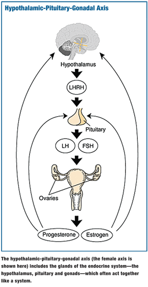
 The endocrine system is comprised of a group of glands found throughout the body. These glands are made up of specialized cells that secrete a substance (such as a hormone) in response to a signal.
The body has two major types of glands: endocrine (which secrete hormones directly into the bloodstream) and exocrine (which secrete substances into a duct that opens onto an external or internal surface of the body).
Endocrine glands release regulatory substances to target organs, whereas exocrine glands secrete substances that are protective and functional.1
The endocrine system provides a network for regulation and integration of all body cells. The main functions of the endocrine system are growth, maturation, metabolism and reproduction. The glands of this system include the pituitary, thyroid, parathyroid, adrenal, gonads, pancreas, thymus and pineal glands.1
Hypothalamic Control
The pituitary gland, also known as the hypophysis, is located directly beneath the base of the brain, just below the optic chiasm.2 It is lodged in a deep depression in the sphenoid bone, known as the sella turcica (saddle-shaped hollow), and is covered by a tough diaphragm (diaphragma sellae).1 The lateral walls of the sella adjoin the cavernous sinus, which contains the internal carotid arteries along with cranial nerves III, IV, V and VI. Inferiorly, the pituitary gland is separated from the sphenoid sinuses by a thin layer of bone.1,3
Although it’s only about the size of a kidney bean, the pituitary gland is one of the most important organs of the body, producing hormones and having reciprocal relations with other endocrine glands. The hormones secreted by the pituitary gland are directly controlled by the hypothalamus.
The pituitary gland also has neural and vascular connections with the brain.2
Hormone Activity
The pituitary gland is made up of two parts: the anterior lobe and the posterior lobe. The anterior lobe, also known as the adenohypophysis, is composed of cells that secrete predominantly protein hormones, including:1,3
• Adrenocorticotropic hormone (ACTH), or corticotrophin, stimulates the adrenal gland (adrenal cortex) to produce glucocorticoids (cortisol).
• Thyroid-stimulating hormone (TSH), or thyrotropin, is a glycoprotein that stimulates the thyroid gland to produce and secrete thyroid hormones (thyroxine).
• Growth hormone (GH), or somatotropin, indirectly promotes growth throughout the body and also controls protein, lipid and carbohydrate metabolism. GH regulates blood nutrient levels. Excessive secretion of GH in young children or adolescents leads to a rare condition known as gigantism. In adulthood, excessive secretion of GH results in the abnormal enlargement of tissues, known as acromegaly.
• Prolactin stimulates the production of milk following delivery in females.
• Follicle stimulating hormone (FSH) stimulates the growth and secretion of ovarian follicles in females and the production of sperm in males.
• Luteinizing hormone (LH) stimulates ovulation and the formation of the corpus luteum. In males, it influences the secretion of testosterone.
LH and FSH are known as gonadotropic hormones because they influence the reproductive system. LH and FSH are essential for reproduction.
The posterior lobe, or neurohypophysis, is an outgrowth of the hypothalamus and is embryonically derived from neural cells. Oxytocin and vasopressin are two hormones that are secreted in the hypothalamus and then released by the posterior pituitary.3
• Oxytocin stimulates the uterus to contract during delivery and helps the uterus to stay contracted after delivery (to prevent hemorrhage). It also stimulates milk ejection.
• Vasopressin (antidiuretic hormone) influences resorption of water by the kidneys and increases blood pressure.1
 Pituitary Tumors
The most common lesion found in the suprasellar area is pituitary adenoma. Pituitary adenomas are slow growing, benign tumors of the pituitary gland. They represent 10% to 15% of all brain tumors.3
Pituitary adenomas are classified as either microadenomas (<10mm in diameter) or macroadenomas (>10mm in diameter). Macroadenomas are frequently found by eye care professionals because they produce visual field defects caused by chiasmal compression.4
A pituitary tumor frequently results in a bitemporal hemianopsia. However, if the optic chiasm is in a prefixed position or the pituitary tumor projects posteriorly, an incongruous homonymoyus hemianopsia may be present because of optic tract involvement.
If the optic chiasm is postfixed or the pituitary tumor projects anteriorly, a compressive optic neuropathy may be present.3,4
Headaches (usually frontal), diplopia, decreased vision, seesaw nystagmus and visual hallucinations are common symptoms of patients who have pituitary tumors.
A prolactinoma is a type of pituitary tumor that produces excess prolactin. Prolactinomas are the most common hormone-secreting pituitary tumors.
A pituitary apoplexy—acute hemorrhagic infarction of the adenoma, resulting in its enlargement—may present with accompanying nausea, vomiting, depression of consciousness and sudden onset opthalmoplegia. Pituitary apoplexy is considered a neurosurgical emergency.3,4
Other diagnoses may mimic pituitary adenoma.4 (See “Differential Diagnosis of a Suprasellar Mass,” right.)
The endocrine system is a complex group of ductless glands that release hormones. The pituitary gland is central to several body functions. It rests in the sella turcica. Primary lesions in this area arise predominantly from the pituitary, hypothalamus, optic nerve, meninges and surrounding mesenchymal structures. n
Thanks to Kelly Malloy, O.D., for her expert consultation and contribution to this article. Coming in July: The Endocrine System: Part II.
1. Rosdahl CB. The Endocrine System. In: Textbook of Basic Nursing. 6th ed. Philadelphia: J.B. Lippincott; 1995:196-205.
2. Fawcett DW. Hypophysis. In: A Textbook of Histology. 11th ed. Philadelphia: W.B Saunders; 1986;479-99.
3. Hendrick AM, Kahook MY, Daoud YJ, Hazin R. Ophthalmic manifestations of endocrine disorders: approaches and medical management. Curr Opin Ophthalmol. 2009 Nov;20(6):495-503.
4. Wormington CM. Pituitary adenoma: diagnosis and management. J Am Optom Assoc. 1989 Dec;60(12):929-35.

