 Q: A 27-year-old white female presented with a two-week history of irritation and foreign body sensation in the left eye. But the slit lamp appearance shows that the eye is white and quiet. What am I dealing with and how should I treat it? Is comanagement with a corneal specialist necessary?
A: “Based on the symptoms and clinical appearance, this patient most likely has Thygeson’s superficial punctate keratitis (TSPK),” says Sheila Morris, O.D., currently a resident at Omni Eye Center, in Atlanta.
As with this patient’s presentation, most individuals with TSPK have a history of irritation, foreign body sensation, photophobia and tearing, Dr. Morris says. These symptoms are typically bilateral and asymmetric, but may be unilateral, as it was in this patient.
Slit lamp examination reveals a white, quiet eye with small, central, corneal epithelial opacities, she says. TSPK lesions are round, granular, white-gray, and stain faintly with fluorescein. A subepithelial haze may be present under these opacities, while the stroma and endothelium are uninvolved.
Differential diagnoses include:
• Herpes simplex virus (HSV) keratitis. “HSV patients typically have a single, red, painful eye, with either a diffuse mild epithelial haze or the standard dendritic ulceration,” Dr. Morris says. “Corneal sensitivity may be reduced in HSV, but not in TSPK.”
• Sterile corneal infiltrates. “Sterile infiltrates are more likely peripheral, and have an intact epithelium,” she says.
• Standard SPK. “The lesions found in standard SPK are much smaller, more diffuse and stain brightly with fluorescein,” she says.
No specific etiology of TSPK has yet been found. “A viral cause has been postulated, but PCR studies have shown no viral involvement,” Dr. Morris says.1 “An immunological component has also been theorized because of TSPK’s clinical response to steroids.”
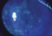
Thygeson’s SPK is notable for small, central epithelial opacities. Photo: Ron Melton, O.D., Randall Thomas, O.D.
Studies have shown a possible genetic link to the HLA-DR3 antigen, which is also associated with Graves’ disease, multiple sclerosis and celiac disease.2 This suggests that the antigen could be affecting the immune status of patients with TSPK.
TSPK usually responds to treatment, but with the disease’s many remissions and exacerbations, it can be an extended, time-consuming process. For mild cases, frequent use of artificial tears may relieve symptoms. But many patients require a mild topical steroid, dosed q.i.d. A gradual taper over weeks to months is necessary to prevent immediate recurrence. If these measures don’t resolve the problem, try a stronger steroid q.i.d.
Monitor patients on steroids every week or every other week to check IOP.
The next step is to try a bandage soft contact lens. “Referral to a corneal specialist is only required in severe cases,” Dr. Morris says.
If studies determine that TSPK does indeed have a viral etiology, then topical antivirals would likely help resolve the signs and symptoms.3 “We did in fact start this patient on Zirgan (ganciclovir gel, Bausch + Lomb) five times a day” Dr. Morris says. “After two weeks, her corneal epithelium was almost completely healed with only a few faint areas of haze remaining. We decreased Zirgan to t.i.d. and added Pred Forte (prednisolone acetate 1%, Allergan) q.i.d., and will see her again in two weeks. Zirgan has been known anecdotally to have an effect on adenoviral conjunctivitis, so it will be interesting to see what happens as we taper it.”
Lastly, educate patients that TSPK is a recurrent disease that may reoccur after several months to many years. “Most importantly, reassure patients that TSPK usually resolves without any long-term effect on vision,” Dr. Morris says.
1. Reinhard T, Roggendorf M, Fengler I, Sundmacher R. PCR for varicella zoster virus genome negative in corneal epithelial cells of patients with Thygeson’s superficial punctate keratitis. Eye. Mar 2004;18(3):304-5.
2. Darrell RW. Thygeson’s superficial punctate keratitis: natural history and association with HLA DR3. Trans Am Ophthalmol Soc. 1981;79:486-516.
3. Nesburn AB, Lowe GH 3rd, Lepoff NJ, Maguen E. Effect of topical trifluridine on Thygeson’s superficial punctate keratitis. Ophthalmology. Oct 1984;91(10):1188-92.
