 If you look at the modern-day optometric practice, you’ll see countless examples of incredible technological devices. From spectral-domain optical coherence tomography to electronic medical records, our offices rapidly are becoming fully automated and computerized. Even our patients are now regularly using smartphones, iPads and all kinds of other electronic gadgets.
It is to the point where the term “manual” is an outdated concept. Yet, one of the most fundamental tools in optometry during the last 80 years has been the completely manual, absolutely non-digital phoropter. But, now that we are beginning to see sweeping technological advances in refraction, is it finally time to replace your “old-school” phoropter?
Out With the Old...
The late Dr. Borish once suggested there were three tests that bother patients the most:
- Tonometry, because of drops and the infamous air burst.
- Visual fields, because they are tedious and difficult for patients.
- Looking through the phoropter, because patients simply don’t like it and can’t easily differentiate the endpoints.
Advanced diagnostic technologies––including rebound tonometers and ultrafast, automated perimeters––have widely addressed patients’ concerns associated with the first two tests. Only the manual phoropter, as we know it, has yet to be reborn into the 21st century. In order to replace a mainstay tool like the manual phoropter, however, new instruments, such as wavefront aberrometers, must provide at least equal accuracy and efficiency as well as offer room for potential improvement.
Objective Measurements of Visual Distortion
Measurements of wavefront, point spread function and root mean square (RMS) error may all be used to determine the amount of objective visual distortion (e.g., myopia, hyperopia, astigmatism, halo, glare, etc.) associated with an individual’s eyes.
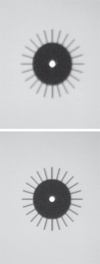
Examples of the targets used during point spread function refraction.
- Wavefront is a measurement of precisely how light rays pass through a patient’s cornea, ocular media and crystalline lens. Wavefront technology analyzes distortions in the patient’s vision and may be used to determine his or her overall refractive error.
- Point spread function is the smallest achievable point of resolution. This measurement can help illustrate true distortions in a patient’s vision.
- RMS error is a mathematical calculation that predicts the overall impact of the patient’s visual distortions. An RMS error greater than 0.43 is considered significant, and could limit a patient’s visual potential with current spectacle lens technologies for myopia, hyperopia and astigmatism in 0.25D steps.1
Wavefront Aberration
One study of 200 patients indicated that an individual’s best subjective focus occurred when the central, aberration-free region of the pupil was maximized.2 Further research showed that objective methods of refraction based on wavefront aberration maps can accurately predict the results of subjective refraction, and actually may be more precise than a manual phoropter.3
Other studies show that eliminating lower-order aberrations, such as myopia, hyperopia and astigmatism, does not necessarily optimize best focus or visual performance.4 So, if correcting higher-order aberrations, defocus and astigmatism will yield the best possible refraction, why haven’t wavefront aberrometers largely replaced manual phoropters?
One reason is that, without a universally accepted metric of image quality, it can be challenging to convert an aberration map into a prescription.3 Additionally, wavefront devices have difficulty properly measuring highly aberrated eyes, such as those found in patients with a history of keratoconus or laser vision correction.5
Then, there is the ultimate subjective variable—we actually see with our brains, not our eyes. So, only the individual patient can truly confirm whether the refractive correction is “better or worse.”
But perhaps the main reason that wavefront aberrometers have not overtaken the phoropter is the risk of over-minusing the prescription.6 One study in particular showed that wavefront analyzer refractions resulted in 0.30D more myopic accommodation (instrument-induced myopia) than required.6
The Technologies
Some of the most widely available devices that can be used to determine objective refraction with great accuracy include: the OPD Scan III (Marco), the KR-1W Wavefront Analyzer (Topcon), iTrace (Tracey Technologies), the Z-View Aberrometer (Ophthonix), the i.Profiler (Carl Zeiss Vision) and the PSF Refractor (VMax Vision). But, can we fully utilize these systems in our practices, given the potential limitations associated with highly aberrated eyes, neural pathways and over-minusing? Or, are these technologies now advanced enough to overcome these shortcomings?
Research has indicated that, while higher-order aberrations influence the amount of sphere and cylinder required to correct vision, subjective refraction can be predicted from the eye’s optics alone by optimizing computed retinal image quality.7 Unfortunately, the data also showed that the range of predictability decreased with greater amounts of higher-order aberrations (e.g., higher RMS numbers).
That said, newer devices, such as the KR-1W Wavefront Analyzer, have been shown to be highly predictable for patients with a history of significant aberrations secondary to dry eye or intraocular lens implantation.8,9
A Look at the PSF Refractor
The PSF Refractor from VMax Vision utilizes
point spread function to optimize a patient’s visual acuity. Point
spread function testing requires a subjective refraction, as opposed to
objective measurements. By presenting point-spread function images above
and below each other, patients generally find it easier to determine
the best refractive choice when compared to Snellen-based manual
refraction.11
The PSF Refractor measures 0.05D changes,
and because it uses point spread images, it automatically corrects for
aberrations in the final prescription. Additionally, because a
point-spread function image will blur when the patient looks beyond the
endpoint––rather than simply appear smaller and darker––the technology
prevents over-minusing. Further, researchers have determined that 95% of
patients achieved identical or better refraction with the device
compared to a manual phoropter.11
Without question, however, these wavefront refraction devices can be used to increase efficiency in your practice. In my experience, for example, the OPD Scan III allows me to perform a 30-second refraction for nearly 75% of my patients. The refraction involves taking the cylinder and axis data as is, and then adding plus to the sphere to ensure that the patient is not overcorrected.
The remaining 25% of patients typically have RMS numbers greater than 0.43 due to irregular cornea shape or previous refractive surgery, and likely would best benefit from manual refraction. (However, it is worth noting that, for highly aberrated eyes, the iTrace has been shown to obtain accurate measurements with high repeatability.10) Regardless, being able to accurately refract 75% of your patients in 30 seconds can offer substantial advantages over solely using a manual phoropter.
Current research suggests that we are now at a point when new technological advances in both wavefront imaging and point spread function testing finally will make it possible for us to replace our phoropters.
These automated devices provide equal or greater accuracy compared to a manual phoropter. Additionally, they will help guide patient decisions by comparing the new refraction directly to the previous one with a single touch of a button (which is difficult to do with a manual phoropter); reduce the rate of repetitive stress injury that many doctors experience; and ultimately make the process of a refraction less stressful on a patient.
It is important to note, however, that eye care providers still will need to assess the final prescription––just as is the case with manual phoropters.
Nonetheless, improved efficiency, more accurate refractive correction orders from patients and the natural patient response to cutting-edge technology should enhance our practices dramatically and finally allow us to replace the oldest piece of equipment in our office.
Dr. Karpecki has served as a paid consultant to Topcon, Marco and VMax Vision, and is on the Speakers Bureau at Carl Zeiss Vision. Neither he nor Dr. Shechtman have any direct financial interest in any of the technologies mentioned.
1. McCormick GJ, Porter J, Cox IG, et al. Higher-order aberrations in eyes with irregular corneas after laser refractive surgery. Ophthalmology. 2005 Oct;112(10):1699-709.
2. Thibos LN, Hong X, Bradley A, et al. Statistical variation of aberration structure and image quality in the normal population of healthy eyes. J Opt Soc Am A Opt Image Sci Vis. 2002 Dec;19(12):2329-48.
3. Thibos LN, Hong X, Bradley A, Applegate RA. Accuracy and precision of objective refraction from wavefront aberrations. J Vis. 2004 Apr 23;4(4):329-51.
4. Applegate RA, Ballentine C, Gross H, et al. Visual acuity as a function of Zernike mode and level of root mean square error. Optom Vis Sci. 2003 Feb;80(2):97-105.
5. Cervino A, Hosking SL, Rai GK, et al. Wavefront analyzers induce instrument myopia. J Refract Surg. 2006 Oct;22(8):795-803.
6. Borisch IM. Clinical Refraction. Vol.1. Chicago: The Professional Press Inc; 1970.
7. Guirao A, Williams DR. A method to predict refractive errors from wave aberration data. Optom Vis Sci. 2003 Jan;80(1):36-42
8. Denoyer A, Rabut G, Baudouin C. Tear film aberration dynamics and vision-related quality of life in patients with dry eye disease. Ophthalmology. 2012 May 14. [Epub ahead of print]
9. Piñero DP, Juan JT, Alió JL. Intrasubject repeatability of internal aberrometry obtained with a new integrated aberrometer. J Refract Surg. 2011 Jul;27(7):509-17. doi: 10.3928/1081597X-20101214-01.
10. Visser N, Berendschot TT, Verbakel F, et al. Evaluation of the comparability and repeatability of four wavefront aberrometers. Invest Ophthalmol Vis Sci. 2011 Mar 10;52(3):1302-11.
11. VMax Vision. Data on file.
