 A 56-year-old Hispanic male presented complaining of central vision loss O.S., which had persisted for the last four months. He reported that he was unable to see faces clearly with his left eye when he covered his right eye. His ocular history is unremarkable; he started wearing reading glasses in his mid-40s and has had no other problems. His medical history is also unremarkable.
On examination, his best-corrected visual acuity was 20/20 O.D. and 20/300 O.S. Extraocular motility testing was normal. His confrontation visual fields were full to careful finger counting O.U., and the pupils were equally round and reactive, with no afferent pupillary defect. Amsler grid testing was normal O.D., but demonstrated a central scotoma involving fixation in the left eye. His anterior segment was unremarkable. Intraocular pressure measured 15mm Hg O.U.
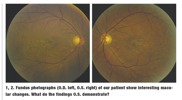
On dilated fundus exam, the vitreous was clear O.U. He had small cups with good rim coloration and perfusion in both eyes. The macula showed obvious changes O.U., as illustrated in figures 1 and 2. Both Stratus (figures 3 and 4) and Cirrus spectral domain OCT (Carl Zeiss Meditech) (figures 5 and 6) were performed, and are available for review. The peripheral retina was normal in both eyes. 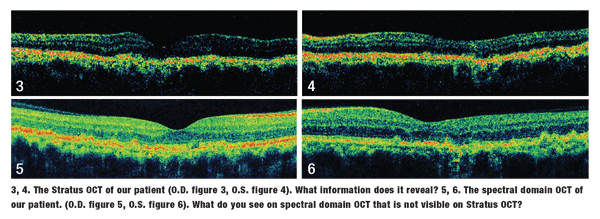
Take the Retina Quiz
1. What do the changes in the maculae represent?
a. Drusen formation.
b. Intraretinal flecks.
c. Intraretinal crystals.
d. Exudate deposits.
2. What does the spectral domain OCT reveal that the Stratus OCT does not?
a. There are no differences between the two scans.
b. Occult choroidal neovascularization (CNV).
c. Loss of the photoreceptors.
d. Intraretinal crystals.
3. What is the correct diagnosis in this case?
a. Non-exudative (dry) age-related macular degeneration (AMD).
b. Exudative (wet) AMD.
c. Retinal angiomatous proliferation (RAP).
d. Biettis crystalline dystrophy.
4. How should this patient be managed?
a. Observation.
b. Vitamins and antioxidants consistent with AREDS recommendations.
c. Intravitreal Lucentis (ranibizumab, Genentech).
d. Intravitreal Avastin (bevacizumab, Genentech).
For answers, see below.
Discussion
The macular changes seen in our patient are nothing more than drusen formation. Drusen are byproducts of metabolic activity from the retinal pigment epithelium (RPE) that lie between the basement membrane of the RPE and Bruchs membrane. Drusen are likely the earliest visible clinical changes associated with AMD.
Macular degeneration is the number one cause of vision loss in people over the age of 50 in the
Even though only 10% of patients with AMD develop the wet form, it is responsible for the overwhelming majority (90%) of AMD-related vision loss. This is due to the development of CNV.
Vision loss may also occur in the dry form, usually from either geographic atrophy of the RPE and choriocapillaris, or from atrophic changes solely within the RPE. In either case, these dry atrophic changes ultimately lead to loss of the photoreceptors.
So, what is wrong with our patient? On careful clinical examination of the maculae O.U., there was no visible fluid or elevation within the retina. Likewise, there was no detectable exudate or subretinal hemorrhage. Do these findings rule out the presence of CNV? Not completely.
Stratus OCT of the maculae O.U. was essentially normal. It showed nothing more than subtle drusenoid changes, which could be seen at the level of the RPE in both eyes. It revealed neither CNV nor any trace of fluid within the retina.
Then, how do we explain the 20/300 acuity O.S.? We could not, so we obtained spectral domain OCT of both maculae. The resolution of Stratus OCT is about 10m, whereas the new Cirrus spectral domain OCT has a sharper resolution, nearly 5m.2 The added resolution allows for much clearer detail of the retinal structures and significantly improved delineation of retinal layers.
At first glance, the spectral domain OCT of our patient did not seem to show much more than what we saw on the Stratus OCT. However, on more careful examination, we were able to see something of significance.
In the image of the right eye that is produced by the spectral domain OCT, almost all of the retinal structures can be seen. The RPE/Bruchs membrane complex is shown as a thick, red or reddish-orange colored line above the choroid and below the sensory retina.
In the right eye, you can clearly observe the irregularities within the RPE layer due to the drusen that we are able to see clinically. Just anterior to the RPE/Bruchs membrane layer, you can also see a thin green line. This line represents the junction of the inner and outer photoreceptor segments. On magnified view, we can clearly see that this line is intact and regular.
We do not observe the same findings in the left eye. Here, we see that the photoreceptor line is not intact, appears very irregular under the fovea, and in fact, is even absent in some areas. This represents loss and disruption of the photoreceptor layer due to atrophy from the AMD, which explains why our patient is experiencing an acuity of 20/300 O.S.
Certainly, a fluorescein angiogram could have been performed; but, we felt that both OCTs were detailed enough to rule out the existence of any CNV.
Though our patient is rather young for a typical diagnosis of AMD, we felt it was accurate, given the clinical findings.
We also believe that this finding could be caused by basal laminar drusen, which, from a histopathological basis, represents the same basic disease process.
We explained these findings to our patient, and recommended that he use vitamin supplements as described by the Age-Related Eye Disease Study (AREDS).
Retina Quiz Answers: 1) a; 2) c; 3) a; 4) b.
1. Klein R, Wang Q, Klein BE, et al. The relationship of age- related maculopathy, cataract, and glaucoma to visual acuity. Invest Ophthalmol Vis Sci 1995 Jan;36(1):182-91.
2. Wojtkowski T, Bajraszewski I, Gorczynska P, et al. Ophthalmic imaging by spectral optical coherence tomography. Am J Ophthalmol 2004 Sep;138(3):412-9.
