VMT is well-defined in the literature, and thanks to advanced optical imaging technology, it is now more easily visualized, diagnosed and monitored.1 Observation and surgical intervention have been described as treatment options.1-4
James’s History
“James,” a 73-year-old man, presented with a complaint of central blur in his right eye that persisted for two days. He reported acute onset, but said that it had remained unchanged since. He denied headaches, diplopia, injury, discomfort and loss of vision.
His medical history included high blood pressure, which he controlled with medication. He was taking Zocor (simvastatin, Merck), lisinopril and a multivitamin.
His ocular history included cataract surgery and LASIK in both eyes. He reported no post-surgical complications.
Diagnostic Data
Best uncorrected distance visual acuity was 20/30- O.D. and 20/25+ O.S. External examination was normal, and there was no evidence of afferent pupillary defect. Refraction found emmetropia O.U.
Intraocular pressure was 16mm Hg O.D. and 14mm Hg O.S. On biomicroscopy, anterior segment structures were normal, and both eyes exhibited mild posterior capsular opacification.
Dilated fundus exam revealed normal optic nerves, vasculature and peripheral retina. Close examination of the maculae revealed an apparent elevation of the right macula with no foveal light reflex. The left macula appeared flat with a normal foveal light reflex.
OCT revealed a posterior vitreous detachment (PVD) with elevation of the macula O.D. (figure 1). There was a full PVD with no macular traction in the left eye.
Diagnosis
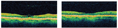
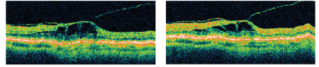
3, 4. At two weeks, James’s
acuity had resolved to 20/20-, but note the free-floating vitreous
cortex (left). At six weeks (right), VA was 20/20, and the cyst had
resolved.
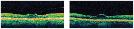
1, 2. At James’s first visit, his
distance visual acuity was 20/30- O.D. (left). One week later (right),
note the changes, even though the VA was unchanged.
Based on the examination findings, James was diagnosed with vitreomacular traction syndrome secondary to a perimacular adhesion of detached vitreous.
Treatment and Follow-Up
We informed James of our findings and educated him about the signs and symptoms of macular hole formation and retinal detachment. He was given an Amsler grid for home monitoring.
At one week, he returned and reported more distortion and blur. But, his VA was stable at 20/30- O.D. OCT showed greater elevation and revealed that the vitreous cortex was still attached to the paramacular area, and there was clearly more intraretinal fluid present (figure 2). There was still no foveal light reflex.
James returned again as scheduled, two weeks after onset of symptoms; he was happy to report his vision was much improved and the metamorphopsia had diminished. Distance acuity was 20/20-, and OCT showed a complete release of macular traction. There was only a small amount of residual intraretinal fluid. The free-floating vitreous cortex could be seen on OCT (figure 3).
An Amsler grid test was performed O.U. for a baseline reference and was recorded as unremarkable O.U. We scheduled James’s next follow-up for a month later to monitor the resolution of the retinal cyst and rule out the possible formation of an epiretinal membrane. At this appointment, he reported no visual effects. Best-corrected visual acuity was 20/20 O.D. and 20/20- O.S. Both eyes were still normal on Amsler grid. On OCT, the PVD was still visible, but there was still no evidence of an epiretinal membrane. The cyst had completely resolved (figure 4).
Kelly’s History
“Kelly,” a 74-year-old woman, presented complaining of blurred vision in her left eye that persisted for almost two months. She reported that it had become blurry gradually, but that it had remained consistent for the past two weeks. She was taking Coreg (carvedilol, GlaxoSmithKline) for high blood pressure, as well as a multivitamin.
Diagnostic Data
At this visit, Kelly’s best-corrected distance visual acuity was 20/20+ O.D. and 20/30- O.S. External examination was normal, and there was no evidence of afferent pupillary defect.
An Amsler grid test revealed central metamorphopsia. Biomiscoscopy revealed normal and healthy anterior segment structures O.U. IOP measured 16mm Hg O.D. and 14mm Hg O.S. Dilated fundus examination revealed normal optic nerves, vasculature and peripheral retina. Examination showed a distinct PVD and an absence of a foveal light reflex in her left eye. OCT confirmed the PVD (figure 5).
Diagnosis
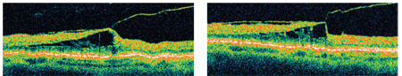
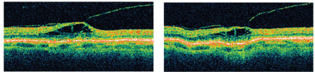
7, 8. At six weeks, the volume of the cyst seemed reduced (left). But, at three months (right), there was still no progression.
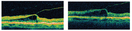
5, 6. At presentation,
Kelly’s left eye showed a PVD and no foveal light reflex (left). Two
weeks later (right), OCT showed an increase in elevation and cyst
formation.
Based on the examination findings, James was diagnosed with vitreomacular traction syndrome secondary to a perimacular adhesion of detached vitreous.
We diagnosed VMT in Kelly’s left eye.
Treatment and Follow-Up
Similar to James, Kelly’s vision was relatively good. So, we chose to monitor her. We gave her an Amsler grid for at-home monitoring, and a follow-up appointment was scheduled for two weeks later.
When she returned, Kelly reported no additional symptoms or change in acuity. But, visual acuity was 20/40+ O.S. OCT of the left macula it showed a slight increase from the previously noted elevation and cyst formation. The vitreous cortex adhesion was still evident (figure 6). We scheduled her to return in one month to monitor the resolution of the retinal cyst and rule out the formation of an epiretinal membrane.
Kelly returned for follow-up and reported no subjective change in acuity and no new or increased symptoms. VA remained 20/20 O.D. and 20/40+ O.S., and the Amsler test results were unchanged. OCT showed that VMT remained, but there appeared to be a slight reduction in the volume of the cyst (figure 7). Eight weeks later, she still reported no change. OCT showed little change in the presentation (figure 8).
Kelly returned one week later concerned that the acuity of her left eye had deteriorated. It measured 20/20 O.D. and 20/50+ O.S. Dilated exam findings remained consistent with previous exams, but OCT showed a slight increase in the cyst volume (figure 9).
|
| 9, 10. Kelly returned with a complaint of visual acuity decrease, attributed to the increase in volume of the cyst after more than three months of monitoring (left). But, at four months (right), symptoms had lessened and her acuity had improved. |
|
|
11, 12. At five months, the cyst had again shrunk somewhat (left). At nine months, her acuity was slightly increased and there was less fluid in the cyst. |
We consulted our institution’s retinal specialist and decided that the general clinical picture of this stubborn VMT and the subsequent VA were reasonably stable. We planned another follow-up in one month and would then consider surgical intervention if there was a significant increase in symptoms, if VA worsened beyond 20/50, or if OCT showed a significant increase in the cyst formation and retinal distortion.
Four weeks later, she had improved. She was less symptomatic, and her acuity was 20/30- O.S. OCT showed a decrease in retinal distortion and cyst volume, so a surgical consult was not considered (figure 10).
This wonderfully compliant patient returned one month later and again reported improvement. Best-corrected acuity was 20/20 O.D. and 20/30+ O.S. OCT showed that the VMT in her left eye was still present, but that there was a slight decrease in cyst volume (figure 11).
As “stubborn yet stable” as her condition was, we believed it reasonable to have her return in six months if there was no deterioration in vision or change in Amsler testing in the meantime.
But, she returned four months later because she was in the area and “just wanted to see how things were going.” She reported that her vision was “as good as it has ever been.” Best-corrected acuity was 20/20 O.D. and 20/25- O.S. Amsler grid testing showed reduction in the degree of metamorphopsia and the size of the area affected. OCT showed that the vitreous cortex still adhered to the macula, but there was yet again less fluid in the cyst (figure 12).
Discussion
Because of its avascular and acellular nature, the vitreous might exhibit a limited number of pathologic changes unless invaded by cells or molecules from surrounding structures. The structural features of the gel, which are apparent in childhood, gradually disappear with time; this process may be accelerated in myopic and aphakic eyes.3,4 As the vitreous degenerates, the collagen fibrils tend to coalesce, and spaces void of any collagen structure develop within the vitreous. With aging, these fluid lacunae may work their way through breaks in the posterior vitreous cortex, which in turn leads to separation between the vitreous cortex and the retina.
The detached vitreous cortex becomes wrinkled and usually separates completely from the retina up to the posterior border of the vitreous base. As that separation progresses through the area of the optic nerve head, a ragged tear in the cortex may be seen: a Weiss ring.5 Often the first subjective sign of a PVD, the Weiss ring may be especially prominent when it has a small amount of glial tissue that detached from the margin of the optic disc. Once the PVD is complete, the flaccid hyaloid surface can be traced to the vitreous base. Complications of PVD depend on the strength and extent of preexisting vitreoretinal adhesions. In most eyes, these are weak enough that PVD presents little threat. However, in up to 10% of all eyes with PVD, that attachment is strong enough that acute PVD may cause retinal tears with vitreous hemorrhage caused by rupture of peripheral blood vessels.6 Unless this kind of tear is treated, the risk of retinal detachment is high.6
Following an acute PVD, the sensory retina is no longer protected by the stable vitreous cortex and can be affected by vitreomacular traction.6 There is no anatomical attachment between the vitreous and the retina at the macula, but during the progression of a PVD, persistent macular adherence can exert tractional force on the macula.2 That force can be centripetal (pulling toward the vitreous), as it was in James’s case, or tangential (pulling nearly parallel to the retinal surface), as it was in Kelly’s case.6 It is not uncommon to see epiretinal membranes and macular holes accompanying or secondary to VMT.3 Clinical characteristics may also include tractional cystoid macular edema, seen in our cases.7
In cases of VMT that do not spontaneously resolve, surgical intervention, including vitrectomy and epimacular vitreous cortex aspiration, show significant visual improvement.3 But, surgical treatments have shown complications of retinal tears, postoperative nuclear sclerosis progression, retinal pigment epitheliopathy and retinal detachment.4 Surgery can also result in such postoperative complications as macular holes or macular atrophy.1 The duration of the vitreomacular change and visual disturbance must be taken into account to find the balance between surgical risk and benefit.
VMT does not behave the same in all cases. While it spontaneously resolved for James in six weeks, Kelly was not so lucky. But, her vision has remained stable and she has remained relatively asymptomatic, so we need only monitor her every few months. She’ll continue home testing, and she’s been told to return to the clinic immediately if sudden changes occur.
Dr. Putnam completed his residency in Primary Care at Wilford Hall Medical Center in 2009. He is currently a staff optometrist at Scott Air Force Base, Ill. Dr. Collins is the Director of the USAF Optometry Residency Program and the Specialty Contact Lens Clinic at Wilford Hall Medical Center, San Antonio, Texas.
1. Yamada N, Kishi S. Tomographic features and surgical outcomes of vitreomacular traction syndrome. Am J Ophthalmol. 2005 Jan;139(1):112-7.
2. Massin P, Erginay A, Haouchine B. Results of surgery of vitreomacular traction syndromes. J French Ophthalmol.1997;20(7):539-47.
3. Koerner F, Garweg J. Diseases of the vitreomacular interface. Klin Monatsbl Augenheilkd. 1999 May;214(5):305-10.
4. Petropoulos IK, Stangos AA, Brozou CG. Vitrectomy for VMT. Klin Monatsbl Augenheilkd, 2003 Mar;220(3):122-6.
5. Spalton D, Hitchings R, Hunter P. In: Atlas of Clinical Ophthalmology. 2nd ed. London: Gower Medical Publishing. 1994;12.4-12.5.
6. Kanski JJ. In: Clinical Ophthalmology. 3rd ed. Oxford: Butterworth and Heinemann, 1994:312-6.
7. Johnson MW. Tractional cystoid macular edema: a subtle variant of VMT. Am J Ophthalmol. 2005 Aug;140(2):184-92.
