Q: I see asymptomatic patients all the time with what appear to be benign choroidal nevi. Do I need to comanage these patients with a retinal specialist?
A: As long as you have the ability to take fundus photographs and are able to compare them, primary care optometrists can follow most of these cases, says Mohammad Rafieetary, O.D., an associate at the Charles Retina Institute, in Memphis, Tenn.
Choroidal nevus is a posterior segment lesion thats quite common, says Dr. Rafieetary. Clinical, post-surgical and postmortem population studies estimate the prevalence anywhere from 0.2% to 32%, he says. But most of these lesions are probably never noticed. Some studies suggest that two-thirds of choroidal nevi go undetected during examination, even with clear media, he says.
So, how are they found?
Choroidal nevi are best detected by indirect ophthalmoscopy with a 20D lens, Dr. Rafieetary says. During this examination, the entire lesion can be seen and compared with the surrounding tissue.
These lesions usually appear flat or slightly elevated, slate gray, and without sharp demarcated borders. They vary in size from 1/3 disc diopter (DD) to 7 DD in basal dimension, and no more than 2mm in thickness.
One of the most common secondary changes in the tissue is the observation of overlying drusen, noted in more than 50% of cases, which is suggestive of longevity, Dr. Rafieetary says. Choroidal neovascular membrane is a rare complication of choroidal nevi.
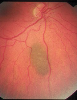 |
| Choroidal nevus with overlying drusen. |
Q: What is the natural history of a nevus? Do they ever turn into melanomas?
A: The primary concern with detecting and following patients who have choroidal nevi is their conversion to malignant melanomas and subsequently metastasis, Dr. Rafieetary says. Its possible that all choroidal melanomas start out as choroidal nevi.
Metastatic disease can occur in choroidal melanomas as small as 3mm in basal dimension and 1.5mm in thickness, he says. Clinical studies show that metastasis occurs within five years in 16% of patients who have small choroidal melanomas (<4mm thickness), as compared with 32% in medium size lesions (4-8mm thickness), and 53% in large lesions (>8mm thickness).1
Yearly follow-up with fundus photography is usually enough to follow patients who have benign lesions, Dr. Rafieetary says. I take an old slide and the new slide, overlie them together and make sure the lesions correspond from slide to slide over time, he says.
While annual fundus photography is the best way to follow these lesions, additional evaluation is needed if there is a change in their size or appearance.
|
How To Find Small Ocular Melanoma |
|
To help in managing or referring patients with choroidal nevi, Dr. Rafieetary uses the mnemonic TFSOMTo Find Small Ocular Melanoma:
Thickness: lesions >2mm
Fluid: any subretinal fluid (suggestive of serous retinal detachment)
Symptoms: photopsia, vision loss
Orange pigment overlying the lesion
Margin touching optic nerve head
None of these factors = 3% risk of a nevus converting to melanoma in five years.
One of these factors = 8% risk of conversion in five years.
Two or more factors = 50% risk of conversion in five years.
For any changes noted during the course of follow-up, refer the patient to a retinal practice or an ocular oncology service. |
Fluorescein angiography, ultrasound and, more recently, optical coherence tomography (OCT) can be used to evaluate these lesions if something appears suspicious, says vitreoretinal specialist Steve Charles, M.D., owner of the Charles Retina Institute. Fluorescein angiography is used to look at abnormal circulation. Ultrasound and OCT are used to measure the height of the lesion and to detect serous detachments.
Any change to the nevus dictates evaluation, Dr. Rafieetary says. He uses a mnemonic to aid in the examination of these lesions. (See How To Find Small Ocular Melanoma, at left.)
Any lesions greater than 3DD in size, any elevation, or any changes noted during the course of follow-up should prompt you to refer the patient to a retinal practice or an ocular oncology service.
Melanomas are typically treated with either radiation or laser, Dr. Rafieetary says, and the success rate is fairly good. The main issue, he says, is to detect the lesion before metastasis.
But, before the lesion even converts to a melanoma, be sure to stress to the patient the importance of regular follow-up. By comparison, a patient who has a similar kind of lesion on the skin could watch it herself and be instructed on when to return to the doctor if it changes. But when its inside the eye, the patient has to come back to the eye care provider to have a look at this, Dr. Rafieetary says.
So, you have to drive home that point that this is not going to cause the patient any symptoms in most cases even if there is some change, and the early change is usually detected by the doctor, he says.
1. Shields CL, Demirci H, Materin MA, et al. Clinical factors in the identification of small choroidal melanoma. Can J Ophthalmol 2004 Jun;39(4):351-7.
|
Ask Dr. Ajamian . . . |
| Do you have a comanagement question or conundrum? If so, ask Dr. Ajamian. Send your question c/o jmurphy@jobson.com, or fax it to (610) 492-1031. |
Vol. No: 141:12Issue:
12/15/04