A 20-year-old Hispanic female presented with blurred vision in her left eye that had persisted for the past two or three months. Her right eye was fine, and she had never worn glasses. She denied any precipitating factors, such as trauma or injury. Her medical history was unremarkable. She grew up in Nicaragua and migrated to South Florida approximately two years ago.

On examination, her best-corrected visual acuity measured 20/20 O.D. and 20/200 O.S. Confrontation visual fields were full to careful finger counting O.U. Pupils were equally round and reactive, with no afferent pupillary defect. Amsler grid testing was normal in her right eye, but showed a central scotoma involving fixation in her left eye. Ocular motility testing was normal. The anterior segment was unremarkable in both eyes. Her intraocular pressure measured 15mm Hg O.U.
Dilated fundus exam revealed a clear vitreous O.U. The right eye was completely normal. The left eye showed a small cup with good rim coloration and perfusion. We noted changes in the left macula (figure 1). We also ordered a spectral domain OCT (SD-OCT) scan of her left eye (figure 2).
Take the Retina Quiz
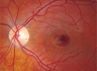
1. Fundus photo of our patient’s left eye. Notice the changes in the macula.
1. How do you account for the fundus changes seen in macula of her left eye?
a. Intraretinal hemorrhage.
b. Choroidal neovascularization (CNV).
c. Hemorrhagic pigment epithelial detachment.
d. Retinal pigment epithelium (RPE) hyperplasia.
2. What is the etiology?
a. Idiopathic.
b. Inflammatory.
c. Infectious.
d. Vascular.
3. What is the correct diagnosis?
a. Valsalva retinopathy.
b. Choroidal nevi with subretinal hemorrhage.
c. Histoplasmosis.
d. Idiopathic CNV.
4. Which statement best describes the clinical presentation and OCT?
a. There is an hemorrhagic pigment epithelial detachment (PED).
b. There is a CNV above the level of the RPE (type II).
c. The OCT shows a suspicious choroidal lesion, but an intact RPE and Bruch’s membrane.
d. This is primary an inner
retinal lesion, because the photoreceptor integrity line (PIL) is essentially intact.
5. How should this patient be managed?
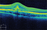
2. The SD-OCT of her left eye is quite revealing. How do you interpret this scan?
a. Observation.
b. Plaque radiation.
c. Intravitreal Avastin (bevacizumab, Genentech) injection.
d. Intravitreal Kenalog (triamcinolone acetonide, Bristol-Myers Squibb) injection.
Retina Quiz Answers: 1) b; 2) a; 3) d; 4) b; 5) c.
Discussion
Our patient has a large subretinal hemorrhage involving the macula of her left eye. There is also a more prominent crescent-shaped, pigmented lesion that can be viewed slightly inferiorly. This represents a CNV that has grown from the choroid, through Bruch’s membrane, and on top of the RP––but below the sensory retina. CNV that grow in this fashion have been categorized has being type II, versus type I CNV that grow below the RPE and are seen in patients with AMD. Type II CNV are more commonly seen in younger patients and are associated with various diseases, such as pathologic high myopia and histoplasmosis, as well as with conditions that cause angioid streaks, such as pseudoxanthoma elasticum (PXE).
Because type II CNV grow above the RPE, they are usually more visible on clinical exam than CNV that are associated with AMD––where the RPE visually obscures them, except on fluorescein angiography (FA) or OCT.
The SD-OCT scan is also helpful in understanding what’s going on with this patient, as well as revealing the anatomical nature of the CNV. Her left macula and fovea exhibit an obvious dome-shaped elevation. More importantly, below the surface, there is a denser, more reflective lesion that is almost triangular shaped with a wider base at the level of the RPE; this is likely the CNV complex. It has caused a significant amount of disruption in the PIL; however, the RPE is largely intact, except for a small area centrally where the RPE is absent. This represents the origin of where the CNV has grown from the choroid, through the RPE, and into the sensory retina.
Undoubtedly, there is also hemorrhage that is within or below the sensory retina.
Why does our patient have a CNV? The most common causes of CNV include AMD, ocular histoplasmosis and pathologic myopia. Other less common causes that can result in CNV include angioids streaks (from PXE), choroidal rupture and post-inflammatory disease, among others.
So, what’s wrong with our patient? She is not myopic, does not have any histo spots and exhibits no signs of previous inflammatory disease. In addition, there is no indication that she had trauma. All told, we concluded that her CNV is idiopathic in nature.
Idiopathic CNV is not so unusual. In fact, it is the fourth most common cause of CNV and was one of the arms of the National Eye Institute’s Macular Photocoagulation Study (MPS). Idiopathic CNV behaves like any other disease that results in CNV.
If left untreated, they can continue to grow and bleed, and eventually result in loss of central vision due to scarring. Treatment options are the same as any other condition that results in CNV, including anti-VEGF therapy.
We explained the examination results to our patient. She received an intravitreal injection of Avastin later that week. We will continue to follow her for visual improvement.
