 |
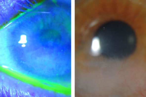
| Changes in cell morphology in both the central cornea and limbal areas may signal LSCD. Click image to enlarge. |
Limbal stem cells (LSCs), located in the limbus of the eye, serve to protect the cornea and help maintain homeostasis of the epithelium, which is vital for visual function. If these cells are not functioning properly as a result of genetic mutation, inflammation or trauma, the culprit is likely LSC deficiency (LSCD), characterized by clinical findings such as stippling or granular fluorescein staining of the metaplastic/conjunctival epithelium.
A useful diagnostic tool for LSCD is in vivo laser scanning confocal microscopy (IVCM), which can help determine disease severity through evaluating central corneal parameters such as basal cell density, epithelial thickness and subbasal corneal nerve fiber length density. In addition to these, a recent study found that assessment of basal epithelial cell morphology (CM) could also help inform LSCD severity, as the condition has been observed to cause changes in CM.
The prospective, cross-sectional comparative study included 168 eyes with LSCD and 63 normal eyes. The researchers performed IVCM on the central cornea and four limbal areas of each eye (superior, inferior, nasal and temporal). A scoring system was developed to assess CM based on basal epithelial cell phenotypes graded from 0 (normal) to 3 (severe morphologic alterations), after which the result was compared with the clinical score, mean best-corrected visual acuity (BCVA) and IVCM parameters.
Compared with the control group of normal eyes, those with LSCD were found to have significantly higher mean CM scores in both the central cornea (1.8 vs. 0.5) and limbal areas (1.6 vs. 1.3). The clinical score correlated positively with the CM score in the central cornea, which correlated negatively with BCVA. All other measured in vivo parameters correlated positively with CM scores.
“CM is an additional biomarker that can be used to confirm the diagnosis and classify the severity of LSCD,” the researchers concluded. “Changes in epithelial CM observed using IVCM included the number of cell layers, cell size and degree of reflectivity of the nucleus and cell-cell junction.”
This newly recognized IVCM parameter can be added to your diagnostic toolbox when examining patients with suspected LSCD. The CM score correlates with the severity of the deficiency and helps inform clinical decision making, the researchers noted.
Bonnet C, Chauhan T, Luna EE, et al. Cell morphology as an in vivo parameter for the diagnosis of limbal stem cell deficiency. Cornea. December 23, 2021. [Epub ahead of print]. |