 |
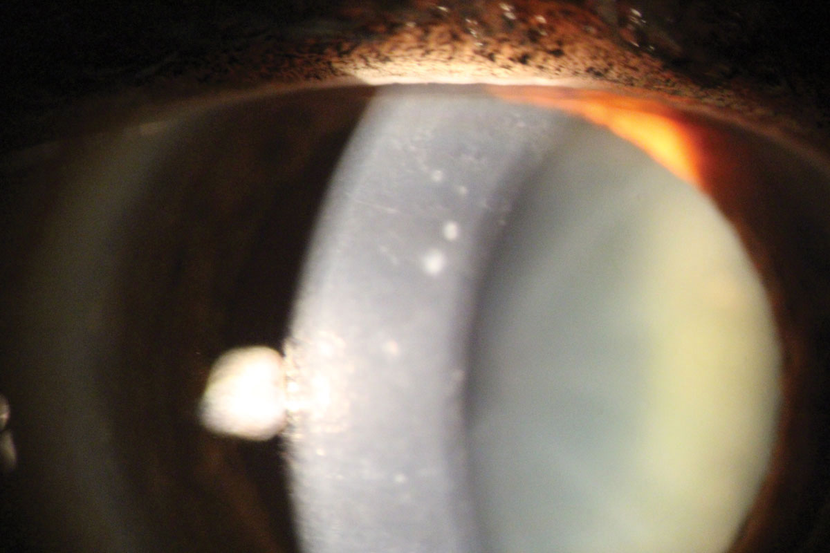
| Among kids, PPCD tends to be unilateral more often than bilateral. Photo: Mitch Ibach, OD and Larae Zimprich, OD |
Posterior polymorphous corneal dystrophy (PPCD) is a rare, slowly progressive autosomal dominant condition distinguished by diffuse, sheet-like vesicular or placoid lesions mostly affecting the posterior layers of the cornea. The majority of patients with PPCD are asymptomatic. Especially in the pediatric population, the condition may go undiagnosed until it leads to visual impairment or other negative outcomes. To better understand its clinical presentation, specifically in children, researchers recently performed a chart review describing the clinical features, topographic and specular microscopy findings and visual outcomes in a group of pediatric patients with PPCD.
Data was collected for 19 patients (27 eyes) who were a mean age of 8.5 years at the time of diagnosis and were followed for a mean of 5.3 years. Researchers reviewed the following: slit lamp findings, cycloplegic refraction, best-corrected visual acuity, central corneal thickness and specular microscopy and corneal topography findings.
While most adult cases of PPCD are bilateral, the results of this study found that the reverse is true of pediatric cases, which supports the findings of previous studies. The dystrophy presented unilaterally in 11 patients and bilaterally in eight. In unilateral cases, differences were detected between affected and unaffected eyes in endothelial cell density, coefficient variation and hexagonality. The mean best-corrected visual acuity at initial presentation was slightly worse in affected eyes than in unaffected eyes (0.8 and 0.9, respectively).
“Focal areas of vesicular or scalloped endothelial lesions were found in 11 eyes, and a band-like pattern was found in 16 eyes,” the study authors wrote. “Seven of the 11 affected eyes in the unilateral group and 13 of the 16 affected eyes in the bilateral group demonstrated progressive endothelial cell loss.”
For 12 eyes, corneal topography was obtained at initial presentation. The mean K-reading for these patients was 43.9D, with significant differences found between mean K-readings of PPCD-affected eyes of patients with unilateral (45.0D) and bilateral disease (41.7D), as well as between PPCD-affected eyes (45.0D) and unaffected eyes (44.2D) of unilateral cases. The mean astigmatism in PPCD-affected eyes was 1.3D at initial presentation and 1.9D at the final follow-up.
Another interesting finding from this dataset was the high prevalence of amblyopia among the studied pediatric patients with PPCD. Seven of 27 eyes had the condition, five of which resolved at least partially with treatment.
“PPCD can present early in children with astigmatism and anisometropic amblyopia,” the researchers concluded. “A careful slit lamp examination for children presenting with anisoastigmatism is necessary to diagnose PPCD. Such patients should be followed up regularly with cycloplegic retinoscopy to prevent and treat refractive amblyopia if present.”
Elhusseiny AM, Saeed HN. Posterior polymorphous corneal dystrophy in a pediatric population. Cornea. June 22, 2021. [Epub ahead of print]. |