
|

|
Anterior stromal puncture (ASP), first described as a treatment for recurrent corneal erosion in 1986, improves epithelial adherence by inducing scar tissue formation between the epithelium and the anterior stroma.1 Researchers initially suggested that ASP should indent the cornea by one-half of its total register; however, a subsequent report indicated that an insertion depth of just 0.1mm was adequate.2
ASP may be performed between erosive episodes or during active erosion, without the need for debridement (although debridement can be performed prior). The optimal candidate for anterior stromal puncture presents with a persistent area of erosion associated with previous trauma or minimal anterior basement membrane dystrophy.
A single ASP procedure is effective approximately 80% of the time. Surgical failures generally occur when the treatment area is too small to address all active erosions.
|
|
|
For a video of the ASP procedure, click
here.
|
Your Role
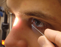
When discussing the procedure with the patient, the term “epithelial reinforcement” may sound more appealing––and yield less anxiety––than “stromal puncture.” After informed consent is obtained, instill several drops of a topical antibiotic and an NSAID. A lid speculum can be inserted, depending on patient cooperation, but typically is not necessary. Instill a few drops of topical anesthetic and place the patient at the slit lamp, instructing them to use the forehead rest at all times. Additionally, an intraocular pressure measurement prior to the procedure will establish a baseline for comparison, and will help you determine if perforation was unsuccessful.
| |
|
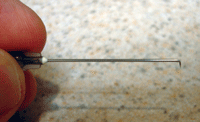
1. A properly bent needle.
|
The technique involves bending the tip of a 22- to 27-gauge needle 90°. The bend at the needle tip should just be long enough to penetrate Bowman’s membrane (i.e., 0.2mm), but not so long that it can enter the anterior chamber (figure 1). The needle should be held tangential to the corneal plane, while the bend should be perpendicular to the corneal surface (figure 2).
Once the needle is prepared, gently indent the affected epithelium. Be sure to create enough punctures to cover the desired area, as well as a 1mm-diameter perimeter around the treatment zone. Place sufficient pressure so that resistance can be felt against the stroma, at approximately 5% to 10% stromal depth. Place the punctures 0.5mm to 1.0mm apart from one another.
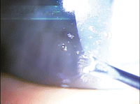
Performing the procedure with fluorescein stain under cobalt blue light will allow bubbles to be visualized. Subepithelial bubbles will be round, while triangular intrastromal air bubbles will indicate that the needle has sufficiently reached the desired stromal depth. Immediately following the procedure, instill a drop of topical antibiotic and a bandage lens. Then, discharge the patient with instructions to use a combination of topical antibiotics and steroids QID for one week.
|
|
|
2. ASP is performed tangential to the corneal plane.
|
Although uncommon, the potential risks with ASP include corneal perforation, corneal scarring, changes in refractive power and topographic irregularities.2
ASP is simple, cost-effective procedure that, aside from the needle, doesn’t require special equipment. No chemicals are used, and because the epithelium remains relatively intact, patients heal very quickly and experience little discomfort.
Dr. Colatrella is the owner and medical director of PineCone Vision Center of Sartell, Minn.
1. McLean EN, MacRae SM, Rich LE. Recurrent Erosion: treatment by anterior stromal puncture. Ophthalmology. 1986 Jun;93(6):784-8.
2. Rubinfeld RS, Laibson PR, Cohen EJ, et al. Anterior stromal puncture for recurrent erosion: further experience and new instrumentation. Ophthalmic Surg. 1990 May;21(5):318-26.
