Supplement to
| NEWS |
|
Check out more articles from the September issue of RCCL here! |
My Perspective: The Myopia Menace Practice Progress: Meet the Candidates Pharma Science & Practice: Pregnant Pause GP Expert: A Lens for Every Eye Out of the Box: Blinded By The Light? Features: Critical Measurements to Improve Scleral Lens Fitting GPs: Are You Making Good Use of Your Diagnostic Fitting Sets? Making The Case for Custom Lenses Options Abound: A 2015 Report on Custom Contact Lenses
|
| Have a topic you want to see discussed? Want to comment on an article? We want to hear from you! Contact us with your feedback and suggestions. |
20 Pearls on Custom Contact Lenses The contact lens industry has nearly perfected mass production of stock lenses—and lately, custom specialty lenses like sclerals, multifocals and orthokeratology lenses are beginning to flourish. With so many options, it’s hard to know what to choose and how to use it. To help, the September issue of Review of Cornea & Contact Lenses is devoted to improving your understanding of the different custom contact lenses available today. Below are 20 pearls shared by experts in the field. For more detail, click through to read the original articles for each. |
|
| 1. As the simplest way to fit GP lenses, empirical fitting saves chair time; however, success using the first lens is only about 40% when employing K readings and refractive measurements. Use topographical software, Sim K information and corneal e-values or asphericity to help make the lens design more accurate. More
| |
| 2. As a general rule, spherical GPs can be used when refractive astigmatism equals corneal astigmatism of less than 2.5D. Any residual astigmatism can be applied to the front surface; however, in cases of higher corneal astigmatism, back-surface or bi-toric GPs should be used. More
|
|
|
4. Try fitting presbyopic patients looking for crisp, uninterrupted vision at both distance and near with translating GP multifocals to avoid inherent limitations of simultaneous vision designs. Pay attention to lower lid anatomy to ensure the lens rests appropriately on the lid and translates upwards as needed. More
| |
| 5. After using a diagnostic lens set, rub and rinse each lens with a cleaner, then disinfect it, blot it and store it dry. When it’s time to use the set again, the lenses should be recleaned, rinsed and applied with a wetting or conditioning solution. More
| |
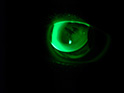
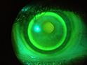
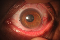
| 6. You can use OCT to evaluate scleral edge profile during fitting: a lens that is too flat will demonstrate edge lift on OCT, which can lead to debris buildup and subsequent fogging of vision, while a lens that is too tight can “dig in” to the conjunctival-scleral complex and lead to discomfort and redness. More
|
|
|
|
| 8. GP multifocals are a good option for high astigmats with presbyopia; these lenses can be fit empirically and typically provide the greatest range of clear vision. Note: some lens movement is typical, but significant movement can impede visual function. More
| |
| 9. When fitting soft toric multifocals for high astigmats with presbyopia, fit the toric aspect first and adjust for rotation and instability before fitting the multifocal aspect. More
| |
| 10. Many GP lens patients experience excessive lacrimation and foreign body sensation as a result of initial lens placement. Use proparacaine during the initial diagnostic lens fitting to numb the eye. More
| |
| 11. Measuring pupil size in both photopic and scotopic conditions can help determine initial zone sizes when fitting either GP or custom soft multifocals. Keep in mind that add power may vary depending on the patient’s most desired working distance. More | |
|
|
| 13. When fitting presbyopes with custom lenses, consider the patient’s desired working distance when making the choice between two lenses. If higher add powers are required, many GP multifocals have adds greater than 3.0D. Some custom labs can also manufacture lenses with an add power up to 4.0D. More
| |
| 14. The introduction of front surface aspheric multifocal or aspheric-concentric combination designs, capable of providing high add powers, has made GP multifocals the “go-to” lenses for presbyopes interested in clearer vision. More
| |
| 15. Sagittal depth of the cornea (influenced by corneal diameter) may play a significant role in the lens/cornea fitting relationship. It’s measured by horizontal visible iris diameter (HVID). Lens base curves and diameter should be manipulated for patients with an irregular HVID measurement to prevent lens decentration. More
| |
| 16. Assess the upper eyelid position when selecting an initial lens diameter for a GP lens. Generally, if the lid is positioned at or near the upper limbus, a lid-attached fit can be achieved with a diameter of 9.4mm or greater. An interpalpebral fit may require a smaller, steeper lens with a diameter of 9.2mm or less. More
| |
| |
| 18. Consider using a specialized diagnostic fitting set for patients with keratoconus, pellucid marginal degeneration and patients who wear bifocal lenses due to variations in pupil size and GP lens designs. More | |
|
| |
|
19. Employ HVID to determine appropriate lens diameter for GP lens stabilization and centration—an inadequate amount can lead to excessive lens movement and discomfort and subsequent patient dropout. More
| |
| 20. Problems with first- and second-generation hybrid designs include limited oxygen transmission, tearing of the junction between the center GP and soft skirt, and a tight fitting relationship. More recent designs have since addressed these issues. More | |
|
| |
| |