Corneal Pain Presentations: Causes and Interventions
Get up to speed on the basis and manifestations of neuropathic as well as neurotrophic changes.
Advances in Endothelial Surgery: An Update for ODs
Over the last few years, there have been radical changes in the treatments for corneal endothelial disease that have impacted the care optometrists provide to their patients. As our treatments evolve and become more effective for Fuchs’ dystrophy and other forms of endothelial dysfunction, intervention often occurs much earlier in the disease process. For instance, we sometimes perform Descemet’s membrane endothelial keratoplasty (DMEK) for patients with 20/20 vision in a dark room assessed with our typical high contrast Snellen charts.
Sizing Up Keratoconus: The Roles of Topography and Tomography
If you are like me and regularly find yourself evaluating topographical maps, you have probably had a variety of thoughts. When starting out in an academic setting, you might have asked yourself: What do these values mean? Which numbers should I pay the most attention to? How do I diagnose conditions based on these maps? Which patients need to have a scan performed on their corneas? Eventually in practice, you may be wondering: Which instrument is worth the money? Which data is reliable, and which is misleading? Now that I have the basics, where can I take this data further in my patient care? This article will answer questions that help distinguish how to best use these devices or read the data.
Corneal Cases: Which are Right For You?
A wide array of corneal conditions are encountered on a daily basis in most optometric practices and appropriate diagnosis and management are vital to ensure good vision and promote ocular health. While many can be managed by practicing OD in their offices, a variety will require surgical intervention or at least a consult with a cornea or oculoplastics subspecialist.
Corneal and Allergy Conundrums
Let’s dive into both worlds and explore new treatments.
The News Feed
Optic Nerve Head Structure Affected by Birth Status
Patient Survey Describes Dry Eye Management Habits, Burdens
Study: Epi-off Accelerated CXL Yields Good Results
Heavy Smoking, High BMI Associated With Earlier Onset nAMD
Access to Pediatric Eye Care Severely Lacking Across the US
Statins Decrease Graves’ Ophthalmopathy Risk
Glaucoma Rate 3x Higher in Poor and Minority Populations
Dry Eye Patients Have Worse Sleep Quality
New Punctal Plug Features Tapered End for Easier Insertion
Glaucoma-related Blindness Decreased Between 2000 and 2020
Structural Complications Not Specific to High Myopia
MGD: A Condition with Multiple Disease Pathways?
Thyroid Function-related Hormones a Risk Factor for DR
Parkinson’s Disease Shows Distinct Retinal Thinning
Age of Myopia Onset Shown to Predict High Myopia
Two Parameters Associated with Comfort in Soft CL Wear
New Drug Combination Successful Against Bacterial Keratitis
Retinal Finding Linked to Myocardial Infarction
Study Documents Substantial Racial Disparities in Glaucoma

Look Inside The Current Issue

Features
Advances in Endothelial Surgery: An Update for ODs
Corneal Cases: Which are Right For You?

Building a Top-Flight Staff
Corneal Pain Presentations: Causes and Interventions
Sizing Up Keratoconus: The Roles of Topography and Tomography
Departments

Attack of the Clones

Big Things in Small Packages
Corneal and Allergy Conundrums
Enough Data to Track Glaucoma Patients?
Flashes? Think Beyond the Retina
Hot Topic

The Enemy Within

Twin Tumor Therapies

Upcoming Events


Continuing Education
Corneal Pain Presentations: Causes and Interventions
OCT Beyond the Basics: Unlocking the Power of This Essential Tool
Demystifying the Complement System
The Physical Manifestations of Glaucoma and What They Signify
Additional Publications
-
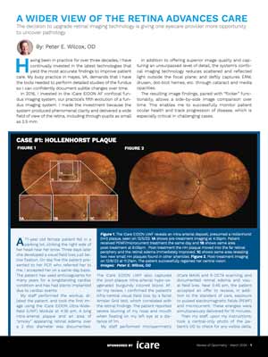
A Wider View Of The Retina Advances Care
The decision to upgrade retinal imaging technology is giving one eyecare provider more opportunity
to uncover pathology.Sponsored by iCare
It’s Time to Talk to Your Patients about Digital Eye Strain
New Developments in Glaucoma
Ophthalmic Product Guide - February 2024
Preservatives in Eye Care: Intrepid Eye Society Consensus Discussion
2024 Conference Planner
Review of Cornea & Contact Lenses

-
Custom vs. Standard Soft Lenses for the Irregular Cornea: How to Choose
Learn which approach works best in this case-based article. -
Corneal Topography: Get to New Heights
See how elevation data reveals the greatest truths about corneo-scleral shape. -
Soft Toric Lenses: Harness This Valuable Practice Opportunity
Experts demystify common misconceptions and offer fitting pearls. -
Wave Hello to Wavefront-Guided Sclerals
These lenses are a great option for those with residual higher-order aberrations but also can be used to create excellent multifocals. -
GP Multifocal Contact Lenses: The 2024 Lineup
Recent design advancements give clinicians even more options to help meet patients’ vision demands. -
Empirical Fitting of GP Lenses
Advanced technology has paved the way for a quite easy and successful approach.
Women In Optometry continues to be published online, with regular updates on practice design, practice success, news, trends and perspectives. Visit womeninoptometry.com.
Job of the Week