 |
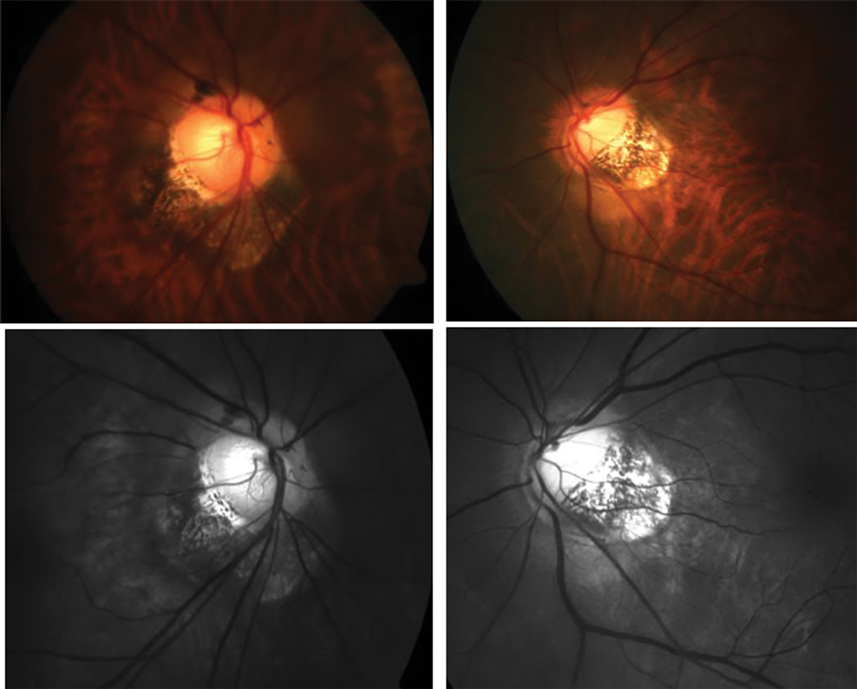
| This study identified several morphological characteristics of the optic nerve head that could predict choroidal thinning and fundus lesions in myopic patients. Photo: Andrew Rouse, OD. Click image to enlarge. |
Certain optic nerve head characteristics may hold clues to myopia pathology, according to a recently published study. Researchers examined the relationship between several of these characteristics and choroidal thickness as thinning of this structure has been found to cause a significant structural change in the course of myopia progression. They identified several risk factors.
The longitudinal study included 323 subjects aged 17 to 30 with healthy myopic eyes. Each participant underwent ocular examinations and measurements for the following parameters: Bruch’s membrane opening (BMO) distance, border length, border tissue angle, peripapillary atrophy, axial length and decreased macular choroidal thickness.
The researchers reported that subjects with longer axial lengths and wider BMO distances were more likely to have thinner choroids in the macular region. They noted that these are significant risk factors. On the other hand, subjects with larger border tissue angles were less likely to develop choroidal thinning, suggesting border tissue angle plays a protective role in choroidal deterioration.
In a basic risk model, the area under the curve—a statistical measure of the correspondence between two variables—for macular choroidal thickness was 0.6284. This value increased to 0.6967, 0.6944 and 0.7383, respectively, after BMO distance, border tissue angle and axial length were added separately to the model. Combining those three parameters resulted in the highest area under the curve: 0.7608.
The researchers concluded that these morphological characteristics of the optic nerve head could serve as early predictors of choroidal thinning and myopia-related fundus lesions in young myopic eyes. “Young myopic patients with [border thickness] configuration change, especially with greater Bruch’s membrane opening distance or small border thickness angle, should be paid more attention and given long-term and regular screening,” they wrote in their paper.
Hu G, Xie J, Shi Y, et al. Morphological characteristics of the optic nerve head and impacts on longitudinal change in macular choroidal thickness during myopia progression. Acta Ophthalmol. May 25, 2022. [Epub ahead of print]. |