 |
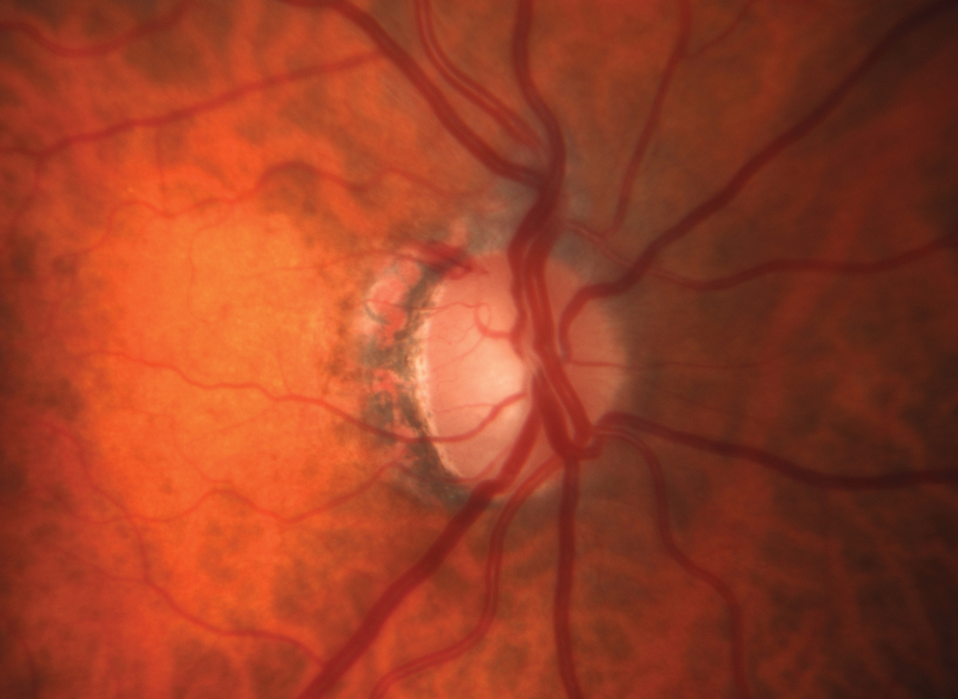
| Hemoglobin levels in the optic nerve head are associated with glaucomatous damage. Click image to enlarge. |
Early detection and treatment of glaucoma is the most effective way to prevent blindness in patients with this highly vision-threatening condition. Optical coherence tomography (OCT) is one of the most effective ways to quantitatively evaluate structural changes in these patients and accurately monitor for progression. However, a study group recently investigated a lower-cost method involving the measurement of hemoglobin (Hb) levels in the optic nerve head (ONH) using automated colorimetric analysis. They found that ONH Hb levels are strongly associated with both structural and functional damage in glaucoma.
The researchers used spectral domain OCT and standard automated perimetry to evaluate the levels of structural and functional damage, and they assessed levels of ONH Hb using automated colorimetric analysis of conventional retinography. Included in the study were patients with primary open-angle glaucoma (n=69) and a control group of healthy individuals (n=54). Using the equipment mentioned above, the following parameters were assessed: ONH Hb levels, glaucoma discriminant function (GDF) index, average peripapillary retinal nerve fiber layer (pRNFL) thickness and visual field mean deviation (VFMD) index values.
All parameters differed significantly between the two groups. In patients in earlier disease stages, more pronounced changes were found in GDF values. The team also found significant nonlinear correlations between GDF and VFMD values, as well as between pRNFL thickness and VFMD. GDF and pRNFL thickness values were found to have a linear correlation.
“While mean pRNFL thickness gradually diminishes as the disease advances, most changes in GDF values were observed in the early stages of the disease (between controls and mild glaucoma patients),” the researchers noted. “Conversely, functional status as determined by the VFMD tends to decay in the latter stages (moderate and advanced glaucoma) of the disease.”
Another recent study compared the diagnostic ability of the Laguna ONhE program with OCT angiography and found that the two perform similarly. The software, which analyzes conventional retinography to estimate Hb levels at the ONH, could be a more feasible, low-cost option for practices that don’t have access to OCT.
“We believe the results of our study, along with the existing literature, support ONH Hb measurement as a viable and accessible tool for assessing structural damage in glaucoma, likely more related to the vascular component,” the researchers concluded.
Tirsi A, Gliagias V, Moehringer J, et al. Pattern electroretinogram parameters are associated with optic nerve morphology in preperimetric glaucoma after adjusting for disc area. J Ophthalmol. October 13, 2021. [Epub ahead of print]. |