 |
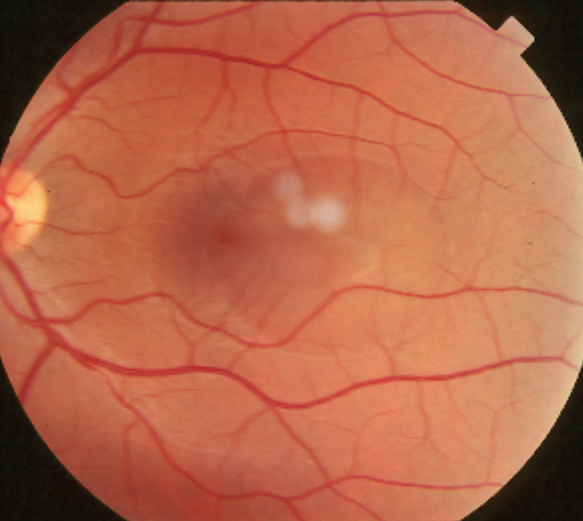
| CSCR could lead to serious retinal problems that threaten vision. Photo: Joseph W. Sowka, OD. Click image to enlarge. |
For most patients with central serous chorioretinopathy (CSCR), the condition will eventually resolve on its own without the need for treatment. However, over time, recurrent or chronic CSCR could cause damage to the retinal pigment epithelium (RPE), resulting in vision-threatening conditions such as RPE atrophy and/or macular neovascularization (MNV), which affects up to one in four patients with CSCR.
One way to check for MNV in this patient population is by evaluating the presence of a “double-layer sign” (DLS). Since most cases of MNV in CSCR are type 1 MNV, indicating that they are located under the RPE resulting in its elevation, this biomarker could help identify such retinal irregularities. A DLS is considered present if a shallow pigment epithelium is at least 1000µm long and has a maximum height of 150µm. However, it’s important to note that once a DLS is observed, OCT-A must be utilized to confirm MNV, since an elevated RPE could also present in patients who don’t have MNV but rather an accumulation of sub-RPE fluid or debris.
Researchers recently set out to test the diagnostic accuracy of DLS and OCT-A in detecting the presence of MNV. They included 163 eyes of 132 patients from an eye hospital in the UK, each with a clinical diagnosis of CSCR. Patients underwent OCT and OCT-A scans, which researchers then reviewed to identify the presence or absence of RPE elevation.
In 91% of eyes, elevated RPE was detected. More than half (58%) of these eyes were found to have DLS. After OCT-A was performed, MNV was confirmed to be present in a third of all eyes.
“Although there was a significant association between the presence of DLS on OCT and the detection of OCT-A evidence of MNV, several cases with neovascularization did not manifest as a DLS,” the study authors noted. “We were able to detect MNV in eyes with an elevated RPE that was as short as 622µm with a height of 412µm. Additionally, the height and length of the RPE elevation were not significantly associated with MNV. Thus, we believe that the size of the RPE elevation in CSCR should not be used to exclude the possibility of the presence of neovascularization.”
The study authors concluded that while a double-layer sign is useful in detecting some cases of MNV, following up by performing an OCT-A scan is an effective way to more accurately confirm that the patient has MNV secondary to CSCR.
Hagag AM, Rasheed R, Chandra S, et al. The diagnostic accuracy of double-layer sign in detection of macular neovascularization secondary to central serous chorioretinopathy. Am J Ophthalmol. October 14, 2021. [Epub ahead of print]. |