
A
weekly e-journal by Art Epstein, OD, FAAO
|
|
Off the Cuff: A Glimpse Into Our Future
Last week I briefly touched on some of the innovations I saw at the Consumer Electronics Show. While the domestic chore-aversive nerd in me is tempted to write about the washing machine that also dried and perfectly folded clothes or the televisions with images so real you could almost walk into them, this week I’ll focus on innovations in eye care.
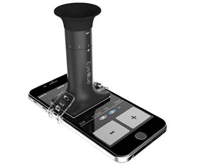
Essentially it combines Shack-Hartmann-based refraction with a smartphone app to provide a user with a reasonably accurate refraction. I’ve tested the unit, and while it may not be as precise as a skilled human refractionist, it’s typically not that far off. What I suspect will be shocking to many is that the app is free and the unit sells for under $30. That’s not a typo. It sells for $29.99 and will soon be on the Home Shopping Network.
EyeQue founder Dr. John Serri is an affable and sharp MIT-trained PhD. We spoke last year and had a lengthy chat last week. His original vision was to create an affordable and accurate means of refraction for the many who don’t have access to vision care. He shared that he found little interest from optometric organizations that he approached. And while he remains open to working with optometry and ophthalmology, at this point, direct sales to consumers has exploded, as evidenced by an exhibit booth several times larger than last year. So, with this “cat out of the bag,” the question becomes, how can we leverage this technology to best benefit our patients as well as our practices? Think fast.
|
||||||


|
||
| Visual Fixation Instability in Multiple Sclerosis Measured Using SLO-OCT | ||||
Precise measurements of visual fixation and its instability were recorded during optical coherence tomography (OCT) as a marker of neural network dysfunction in multiple sclerosis (MS), which could be used to monitor disease progression or response to treatment. A total of 16 MS patients and 26 normal subjects underwent 30 seconds of scanning laser ophthalmoscope (SLO)-based eye tracking during OCT scanning of retinal layer thickness. Study groups consisted of normal eyes, MS eyes without prior optic neuritis (MS wo ON), and MS eyes with prior optic neuritis (MS + ON). Kernel density estimation quantified fixation instability from the distribution of fixation points on the retina. In MS wo ON eyes, fixation instability was compared to other measures of visual and neurologic function. Fixation instability was increased in MS wo ON eyes (0.062 deg2) compared with normal eyes (0.030 deg2). A further increase was seen for MS + ON eyes (0.11 deg2) compared with MS wo ON and normal eyes. Fixation instability correlated weakly with ganglion cell layer (GCL) volume and showed no correlation with low-contrast letter acuity, EDSS score or SDMT score. Fixation instability reflects the integrity of a widespread neural network germane to visual processing and ocular motor control, and is disturbed in MS. Further study of visual fixation, including the contribution of microsaccades to fixation instability, might provide insight into the localization of fixation abnormalities in MS and introduce innovative and easily measured outcomes for monitoring progression and treatment response. |
||||
SOURCE: Mallery RM, Poolman P, Thurtell MJ, et al. visual fixation instability in multiple sclerosis measured using SLO-OCT. Invest Ophthalmol Vis Sci. 2018;59(1):196-201. |
||||
|
|||

| Correlation Between Morphological Characteristics in Spectral-Domain-Optical Coherence Tomography, Different Functional Tests and a Patient's Subjective Handicap in Acute Central Serous Chorioretinopathy | ||||
The purpose of this study was to identify quantitatively measurable morphologic optical coherence tomography (OCT) characteristics in patients with acute episodes of central serous chorioretinopathy (CSC) and evaluate correlations with functional and psychological variables for use in daily clinical practice. Retinal thickness (RT) and the height, area and volume of subretinal fluid (SRF)/pigment epithelium detachments were evaluated using the standardized procedures of the Vienna Reading Center. These morphologic characteristics were compared with functional variables [best-corrected visual acuity (BCVA), contrast sensitivity (CS), retinal sensitivity/microperimetry and fixation stability] and patients' subjective handicap from CSC using the National Eye Institute 25-item Visual Function Questionnaire (NEI VFQ-25).
Data from 39 CSC patients were included in this analysis. Three different SRF height measures showed a high negative correlation with retinal sensitivity within the central 9 degrees, which was also negatively correlated with SRF area and volume. The CS score and fixation stability (fixation points within 2 degrees) showed a moderate negative correlation with SRF height variables. Comparison of the subjective handicap with morphological characteristics in spectral-domain (SD)-OCT showed SRF height had the highest correlation with the subjective problems reported and overall NEI VFQ-25 score. Researchers concluded that SRF height measured in SD-OCT showed the best correlation with functional variables and patients' subjective handicap caused by the disease, and, therefore, seemed to be the best variable to look at for use in daily clinical routine. They added that, even though area and volume also showed a correlation, these could not be as easily measured as height so researchers did not recommend their use in daily clinical practice. |
||||
SOURCE: Gerendas BS, Kroisamer JS, Buehl W, et al. Correlation between morphological characteristics in spectral-domain-optical coherence tomography, different functional tests and a patient's subjective handicap in acute central serous chorioretinopathy. Acta Ophthalmol. 2018; Jan 16. [Epub ahead of print]. |
||||
 |
||
| Autoimmune Retinopathy: A Review | ||||
Autoimmune retinopathy (AIR) is a rare and still poorly understood immune-mediated disease that may cause inflammation from circulating autoantibodies against the retina, authors wrote. The disease may be related to history of autoimmune disease in the patient or a family member, or the presence of neoplastic disease in the individual, can be subdivided into paraneoplastic and non-paraneoplastic AIR, they added. Furthermore, when related to melanoma, it is referred to as MAR, and when related to other cancers, it is called CAR. The exact prevalence of AIR is unknown; it mainly affects older adults, authors explained.
Patients present with bilateral and asymmetric scotomas, photopsias and visual field defects, with rapidly progressive visual loss in late onset, the authors wrote. In the initial stage, fundus examination is unremarkable, and in late stages, evidence can be found of limited retinal epitheliopathy and vascular attenuation, with or without optic disc pallor, associated or not with intraocular inflammation and without evidence of degenerative retinal disease, they continued. A clinical investigation with detailed anamnesis and laboratory tests should be performed to search for an associated neoplasm, and ophthalmologic and complementary examinations such as full-field electroretinogram, optical coherence tomography, visual field and fundus autofluorescence help the diagnosis, they suggested. Author also advised that blood tests to search for autoantibodies should be requested, and management consists of prolonged immunosuppression, which may be combined with antioxidant vitamins. The authors concluded that, in general, prognosis is uncertain, so the disease still needs to be better understood. In conclusion, they wrote, more studies should be performed to improve diagnostic measures and define specific management that could preserve or even restore vision. |
||||
SOURCE: Canamary AM Jr., Takahashi WY, Sallum JMF. Autoimmune retinopathy: A Review. Int J Retina Vitreous. 2018;4:1. |
||||
|
|||

| News & Notes | ||||||||
| FDA Approves B+L Lumify OTC Eye Drop with Low-dose Brimonidine Bausch + Lomb announced the U.S. Food and Drug Administration approved Lumify (brimonidine tartrate ophthalmic solution 0.025%) as the first eye drop developed with low-dose brimonidine tartrate for the treatment of ocular redness. Six clinical studies were conducted in more than 600 subjects to evaluate the safety and efficacy of low-dose brimonidine tartrate in relieving ocular redness, including studies with both pediatric and geriatric subjects. The double-blinded, randomized, placebo-controlled Phase III efficacy study showed that 95% of subjects reported significant symptom improvement at one minute, while 79% of respondents maintained significant redness reduction at eight hours. Lumify will be available for purchase in retailers in the second quarter of 2018. Read more. |
||||||||
Atlantis Scleral Offers Unlimited Exchanges Warranty Policy
|
||||||||
| OCuSOFT Partners with I-MED Pharma to Bring I-Pen to U.S. OCuSOFT announced a new distribution agreement with I-MED Pharma, a biotechnology company, to make the I-Pen Osmolarity System available in the United States. On Dec. 22, 2017, the FDA issued an Acceptance Review Notification for the system’s 510k submission. The distribution partnership will officially begin upon final FDA 510k approval, which is anticipated in the first quarter of 2018. The I-Pen Osmolarity System includes a handheld device that measures the osmolarity of human tears in normal and dry eye disease patients. Read more. |
||||||||
|
||||||||
|
||||||||
|
||||||||
|
||||||||


|
Optometric Physician™ (OP) newsletter is owned and published by Dr. Arthur Epstein. It is distributed by the Review Group, a Division of Jobson Medical Information LLC (JMI), 11 Campus Boulevard, Newtown Square, PA 19073. HOW TO ADVERTISE |