OCT’s versatility provides doctors with a view of the eye that’s helped change the way they practice. |
|
Making a Case for OCT
How imaging technology can change the way you practice.
The speakers in “Understanding and Interpreting the OCT in Retina and Glaucoma” on Thursday morning made a convincing case for the importance of using optical coherence tomography (OCT) in clinical eye care practice for diagnosing, following and managing retina and glaucoma-related conditions.
Noting the first scientific description of OCT in the Journal of Science in 1991, the speakers said OCT is now widely embraced by optometrists and ophthalmologists. Its widespread use is being driven by increased demand for eye care due to an aging population, yielding a growing number of cases involving macular degeneration, diabetic retinopathy, retinal vascular disease and glaucoma.
“What an opportunity for optometry to get involved in medical eye care,” said Mark Dunbar, OD, director of optometric services at Bascom Palmer Eye Institute, “by having an OCT to keep these patients in your chair longer.”
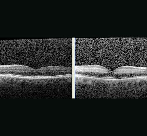
Qualities of a Good OCT
Dr. Dunbar said his experience with various retinal OCT devices and software packages revealed that they all were “very good.” He pointed out that “a B-scan is a B-scan” and that—on both ends of the economic spectrum—all OCT offerings he has tried offer high definition, three- to five-micron images that pick up key features of abnormality and pathology.
Though OCT software programs for glaucoma are unique, Dr. Dunbar said the differences boil down to subjective factors and personal preferences. But he pointed out that all the devices do a “great job” of detecting retinal nerve fiber layer thinning and evaluating the ganglion cell complex. Since the hardware is similar across platforms, software that is patient friendly, competitively priced and fits the needs of clinicians are important purchasing considerations.
When it comes to OCT selection, Jeffry D. Gerson, OD, an optometrist at Grin Eye Care in Leawood, Kan., compared the process to buying different tiers of contact lenses from companies offering “good, better and best” options. “For most patients, the ‘good’ works out just fine,” Dr. Gerson said.
Confidence Factor
During the comprehensive presentation, featuring 245 slides, in which Drs. Dunbar and Gerson gave tips for OCT interpretation of retinal and glaucoma conditions, the speakers urged clinicians not to make OCT interpretation more complicated than it needs to be. With any kind of imaging technology, especially when dealing with anatomy, there can be an “intimidation factor,” they noted. Dr. Dunbar told the story of a friend and colleague who purchased an OCT several years ago.
“He went through all of those insecurities: ‘Is this technology too advanced for me? Am I smart enough to figure it out?’” Dr. Dunbar said. A year later, the friend’s confidence with OCT was high. “It really changed his life and how he practices,” Dr. Dunbar said. “So don’t get caught up in the minutia.”
