In a new study, researchers longitudinally assessed the impact of high-risk structural biomarkers for natural disease progression in non-exudative age-related macular degeneration (AMD) on spatially resolved mesopic and scotopic fundus-controlled perimetry testing. They found that PED and HRF had the greatest prognostic impact on progressive point-wise sensitivity losses.
 |
|
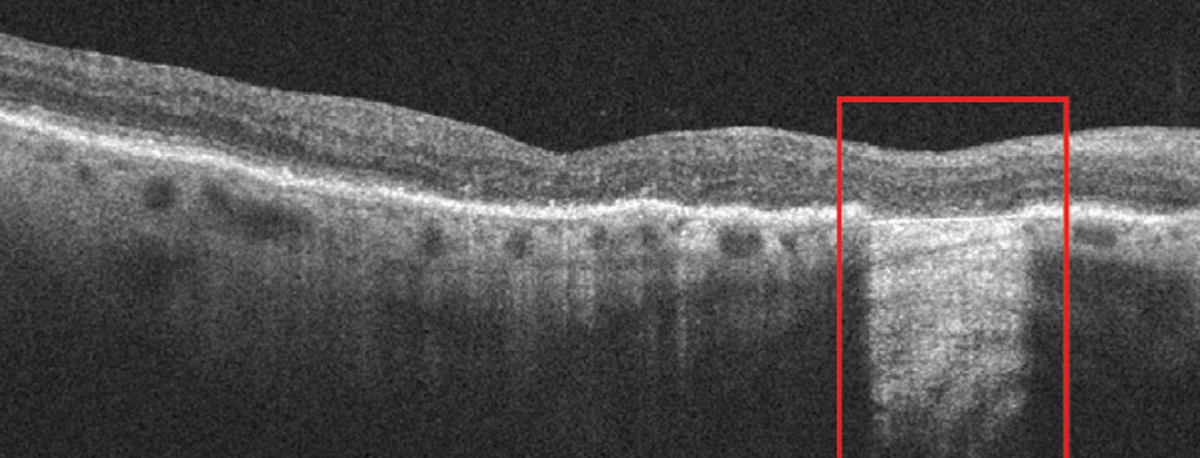
The study found significant progressive functional decline in scotopic visual acuity for patients with cRORA lesions; results with iRORA were more variable. Photo: Anna Bedwell, OD. Click image to enlarge. |
A total of 54 eyes of 49 patients with non-exudative AMD and 27 eyes of 27 healthy controls were included. Multimodal retinal imaging data and fundus-controlled perimetry stimuli points were semi-automatically registered according to landmark correspondences at annual visits over four years. The presence of sub-RPE drusen, subretinal drusenoid deposits, pigment epithelium detachments (PEDs), hyper-reflective foci (HRF), vitelliform lesions, refractile deposits and incomplete RPE and outer retinal atrophy (iRORA) and complete RPE and outer retinal atrophy (cRORA) were graded at each stimulus position and visit.
The greatest impact on mesopic and scotopic retinal sensitivity was detected at retinal locations with the presence of PED and HRF on the non-exudative AMD eyes, whereas the least impairment on retinal function was exhibited in presence of refractile deposits for both sensitivity testings.
“Our results underline previous findings by Kitano et al., revealing a significantly deteriorated mesopic retinal sensitivity at retinal locations with the presence of drusenoid or serous PED lesions without evidence of macular neovascularization,” the researchers wrote in their paper for Investigative Ophthalmology and Visual Science. “Furthermore, our results support findings by Echols et al. reporting a significant correlation between HRF presence and increasing HRF count to a progressive impairment of retinal function, including greater losses of scotopic than mesopic function.”
In non-exudative AMD study eyes, another study recently showed a decreased mesopic retinal sensitivity of -1.422 dB when HRF are present, which was, despite the use of different FCP devices and testing protocols, the authors mentioned, overall higher compared with the mesopic functional impairment of -0.893 dB in HRF presence.
“Interestingly, [in the study mentioned above] and our study, localized mesopic retinal sensitivity was stronger affected by the presence of HRF than sub-RPE drusen, although the overall impact of sub-RPE drusen presence on retinal sensitivity remained significant. These concurring results support the important role of HRF as a high-risk factor for disease progression and functional decline in non-exudative AMD, as reported previously,” the authors stated.
For structural biomarkers of early atrophy development, the greatest sensitivity losses were found for mesopic testing at retinal locations with c-RORA lesions, whereas scotopic retinal sensitivity loss was only significant with the presence of i-RORA lesions. “These results are also in line with previous clinical as well as histological-clinical correlation studies, which suggested that rod dysfunction precedes cone dysfunction in AMD disease, probably owing to a shortening and ongoing degeneration of rod outer segments with disease progression, while cones remain resilient,” the authors explained. “Whether dysfunctional RPE and/or choriocapillaris flow deficits are the primary factor leading to a progressive photoreceptor impairment exceeding normal aging processes is still under debate.”
Regarding the inter-visit analysis, there was a significant progressive functional decline for scotopic testing at cRORA lesions only, whereas for iRORA lesions sensitivity results varied at a low significance level for mesopic testing between follow-up visits.
“These results further suggest that there is a high degree of variability of functional data in patients over time,” the authors concluded. “For a degenerative retinal disease as AMD, where the manifestation of first structural alterations can often take years or decades, even a follow-up period of up to four years with the available FCP device is too short to observe significant changes over time. Moreover, the classification into intermediate AMD according to Ferris et al. may include several different manifestation stages of disease toward atrophy development. Thus, study eyes with only slight structural alterations at baseline visit seem to have no significant changes over time compared with those developing iRORA or cRORA lesions.”
Sabammshausen M, Dongelci S, Vaisband M, et al. Spatially resolved association of structural biomarkers on retinal function in non-exudative age-related macular degeneration over four years. Invest Ophthalmol Vis Sci. April 30, 2024. [Epub ahead of print.] |