Acute unilateral vision loss is typically the first indicator of multiple evanescent white dot syndrome (MEWDS), a particular form of posterior uveitis predominantly affecting women in their third to fourth decade. Given the primacy of visual symptoms to the condition, gaining a deeper understanding of photoreceptor function in MEWDS could help to better characterize the disease.
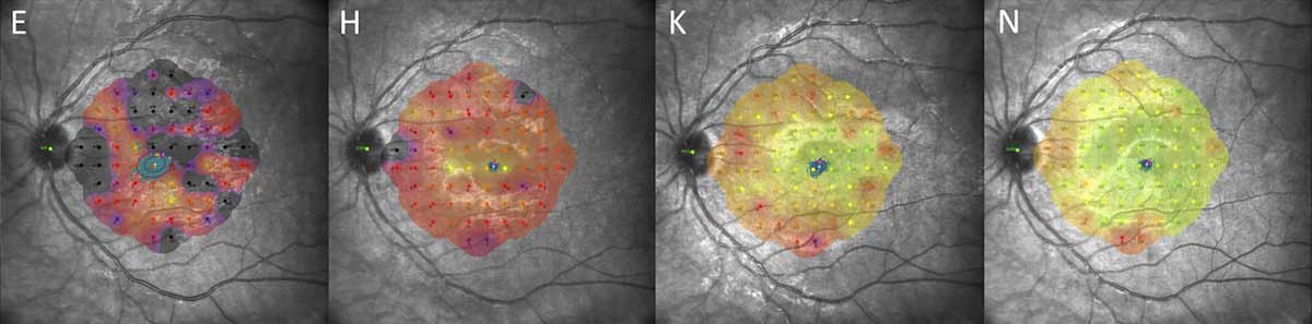 |
| A centrifugal recovery pattern was observed consistently across patients, with faster restoration in the central macula, progression to the extramacular and peripheral regions and culmination around the optic disc. This series from the study (left to right) shows a global reduction in retinal threshold sensitivity by microperimetry at baseline, with darker areas representing deeper scotomata, followed by steady improvements at two weeks, six weeks and three months. Photo: Cicinelli MV, et al. Invest Ophthalmol Vis Sci. 2024;65(4):28. Click image to enlarge. Click image to enlarge. |
Researchers from Milan and London recently investigated structure-function correlations in 14 eyes from 13 MEWDS patients using microperimetry and spectral-domain OCT (SD-OCT). Over a monitored period of median 49.5 days, investigations documented retinal threshold sensitivity (RTS) by means of microperimetry, best-corrected visual acuity, a clinical assessment of foveal granularity and a metric termed photoreceptor reflectivity ratio (PRR), the latter as a measure of photoreceptor integrity. To arrive at PRR values, the team measured light intensity reflected from a band of tissue just above the RPE, approximating the anatomic region of the photoreceptor outer segments. “A lower PRR value indicated diminished reflectivity, suggesting potential damage,” the team explained in their paper for Translational Vision Science & Technology.
Included in the testing were 2,340 microperimetry locations. Early photoreceptor disruption was indicated through transient PPR decrease within 30 days post-presentation; this was followed by a progressive increase, signaling recovery. Eyes with foveal granularity experienced lower foveal sensitivity and increased fixation spread than those without granularity. An increase in RTS was seen over time with a central-to-peripheral gradient of improvement.
More rapid improvement in eyes with worse initial vision was indicated through the interaction between follow-up time and baseline best-corrected visual acuity. There was also a robust, nonlinear association between PRR and RTS across all locations tested.
Placing their findings into context, the authors point out that their study reflects existing literature, spanning a wide spectrum of clinical manifestations. Age and refractive errors also varied widely in this cohort, accounting likely for the difference between those with primary vs. secondary MEWDS. Visual acuity at onset was also found to vary and similarly reflects previous studies.
The use of microperimetry revealed that fixation stability was maintained even when retinal sensitivity was much diminished, suggesting relative central vision preservation in MEWDS. Retinal sensitivity was explored through clinical and demographic associations, with foveal granularity causing detrimental central visual function effects and a wider fixation spread. Since the size of foveal granularity decreased with increasing time from symptom onset, this may be a potential indicator of early disease.
However, detection of vertical hyperreflective lines on imaging studies, which might signify Muller cell activation in MEWDS, did not greatly alter retinal sensitivity measures, the researchers explained in their paper. Subsequently, “indicating that the photoreceptors are taking the greatest brunt of disease impact,” they wrote. On top of this, presence of primary or secondary MEWDS did not affect presenting retinal function or recovery pattern, meaning a stereotyped response is likely present regardless of patients’ characteristics.
The functional recovery that was observed as more pronounced in eyes with worse initial best-corrected visual acuity may suggest a ceiling effect for those with better vision. Along with the distinct centrifugal pattern of visual improvement, the authors relay that “this pattern, alongside the predilection for the peripapillary region, prompts further investigation into the specific vulnerabilities underlying the area around the disc.”
Cicinelli MV, Montesano G, Berni A, et al. Photoreceptor integrity in MEWDS: longitudinal structure-function correlations. Invest Ophthalmol Vis Sci. 2024;65(4):28. |