While intraocular pressure plays a decisive role in both the pathogenesis and treatment of most forms of glaucoma, its influence in normal-tension glaucoma (NTG) is not well understood. Although lowering it has been demonstrated to slow visual field loss progression, even when successful there is still retinal ganglion cell loss with consecutive visual field defects in many patients, calling into question other pathophysiological mechanisms, including cerebrospinal fluid (CSF) dynamics within the subarachnoid space of the optic nerve.
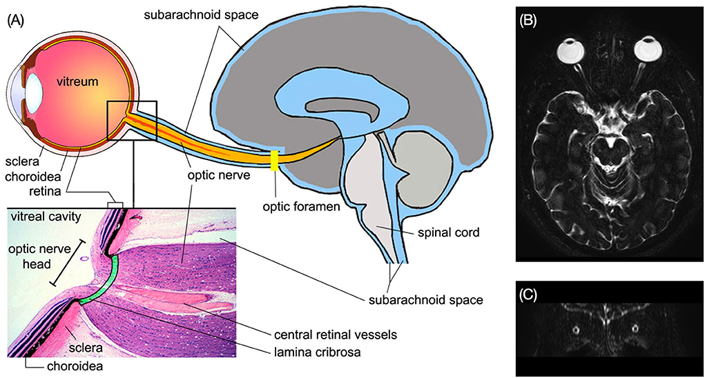 |
| Recent evidence points to CSF dynamics becoming impaired with age, which plays a crucial role in brain health in older individuals. Photo: Sheng J, et al. Front. Neurol., Aug. 14, 2022. Click image to enlarge. |
Researchers from Sweden and Switzerland recently conducted a prospective study aimed to measure CSF flow rates in the subarachnoid space of the ON using noninvasive diffusion-weighted MRI in normal tension glaucoma patients compared with controls. Included were 49 optic nerves of 26 NTG patients and 52 of age-matched controls. Subjects were classified into four groups by age:
- group I, age 50 to 59 (12 eyes)
- group II, age 60 to 69 (16 eyes)
- group III, age 70 to 79 (18 eyes)
- group IV, age 80+ (6 eyes)
Using MRI imaging, the researchers devised a metric they called flow-range ratio (FRR) to estimate flow velocity of moving particles between the frontal lobe and optic nerve subarachnoid spaces. This was calculated for all participants and compared between age categories and between patients vs. controls.
The FRR was significantly reduced in NTG patients vs. controls; however, there was no difference within age categories or the control group, nor was there a difference between subgroups when comparing FRR of NTG patients by age category. The mean FRR by age groups for NTG patients vs. controls, respectively, was: (I) 0.54 and 0.62, (II) 0.56 and 0.63, (III) 0.54 and 0.62 and (IV) 0.61 and 0.61.
CSF flow range was the same in NTG patients for groups I, II and III, but greater in group IV, while controls saw a slight decrease from group I to IV. Characterizing their results in a paper published in the British journal Eye, the authors proposed two possible mechanisms responsible for the decreased CSF flow observed within the subarachnoid space along the ON in NTG patients. One of these is compartmentalization of the ON sheath, while the other explanation could be the effect of low CSF pressure.
As well, the authors mention that adequate CSF flow is vital to maintain proper delivery of neurotrophic factors, electrolytes, neurotransmitters and hormones to neurons, axons and glial cells in the central nervous system. CSF also clears the central nervous system; when it stagnates, this causes accumulation of certain biomarkers of neurodegeneration. Related to glaucoma, CSF samples of NTG patients from a compartmented ON subarachnoid space have demonstrated elevated levels of a multifunctional prostaglandin synthetase; this is indicative of CSF stagnation.
The authors wrote in their paper that, “given the physiological importance of CSF for the integrity of neurons, axons and glial cells, reduced CSF flow dynamics might be part of the underlying neurodegenerative process of NTG.”
Berberat J, Pircher A, Remonda L, Killer HE. Age related cerebrospinal fluid flow dynamics in the subarachnoid space of the optic nerve in patients with normal tension glaucoma, measured by diffusion weighted MRI. Eye (Lond). April 25, 2024. [Epub ahead of print]. |