 |
|
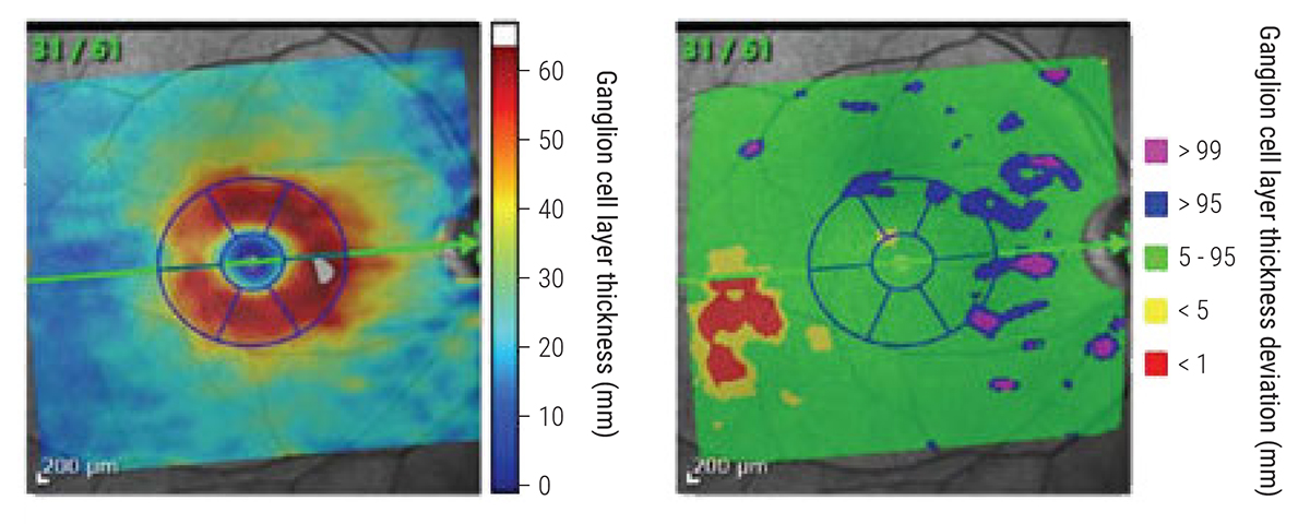
Every advanced glaucoma patient in this study exhibited some extent of ganglion cell loss in the donut-shaped region of the central macula, the health of which is critical for daily tasks such as reading, driving and recognizing faces. Photo: James Fanelli, OD. Click image to enlarge. |
A donut-shaped region of the central macula (±8° from fixation) is critically important for everyday activities, such as reading, driving and facial recognition. This region also contains more than 30% of ganglion cells, despite representing just 2% of the total retinal area. Given glaucoma patients experience a gradual loss in ganglion cells over time as their disease progresses, researchers aimed to determine how much of this damage takes place in this vital region in a recent study published in Journal of Glaucoma.
The retrospective, cross-sectional study involved 94 eyes with advanced glaucoma, defined as a 24-2 mean deviation worse than -12 dB. The researchers measured ganglion cell layer (GCL) thickness on OCT in all patients and used a commercial report to calculate the GCL thickness in six sectors of the donut-shaped GCL region around the fovea. The six sectors were then coded as one of three colors for each eye: green (within normal limits), yellow (≤ 5th, 1st percentile) or red (<1st percentile).
The data showed that all patients in the study had damage to this critical central macular region. In all eyes, at least one sector of the donut was abnormal (red or yellow), while all six sectors were red in 55% of the eyes. Thirty-three eyes, or about one in three, had at least one sector coded as green. The researchers explained in their paper, “While the pattern of donut damage varied widely across these 33 eyes, 61 of the 66 hemiretinas were consistent with a temporal-to-nasal progression of damage within each hemiretina as predicted by our model.”
The authors also pointed out that damage to the TI sector of the donut-shaped region was more extensive than in other sectors. “This is due to the fact that the axons of the ganglion cells in the inferior portion of the macula, including those in the TI sector, enter the inferior vulnerability zone of the disc, which is the region most vulnerable to glaucomatous damage.” On the other hand, the NS sector had the fewest red sectors, which is consistent with prior studies.
These findings highlight the importance of monitoring the progression of damage within the central macular donut region in patients with advanced glaucoma, the authors urge. Additionally, they offer guidance for identifying macular progression, inclusion criteria for clinical trials and selection criteria for future studies aimed at preserving central macular function.
“Given the importance of the central macula to reading, driving and recognizing faces, an emphasis should be placed on saving the GCL donut region,” the researchers concluded.
Sun AJ, Gomide G, Tsamis E, et al. Understanding patterns of preserved retinal ganglion cell layer in advanced glaucoma as seen with OCT. J Glaucoma. April 2024. [Epub ahead of print]. |