 |
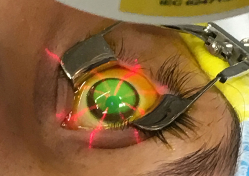
| An accelerated protocol for performing corneal collagen crosslinking, available internationally, showed good long-term safety and stability. Photo: Glaukos. Click image to enlarge. |
Keratoconus can result in devastating outcomes like irreversible vision loss and significant corneal vulnerability, but early diagnosis and the availability of collagen crosslinking (CXL) are greatly improving the long-term picture for many patients. The Dresden protocol for CXL is rather time-consuming and bothersome to patients and practitioners, so international experts have been offering an accelerated version for quite some time, though it has yet to come to the US.
In one cohort of pediatric patients, researchers evaluated the 10-year visual, refractive and tomographic outcomes of those who received epithelium-off accelerated CXL. Included in the investigation were 175 eyes of 97 children (mean age: 14.5). Results of the work were recently published in the British journal Eye.
Uncorrected and corrected distance visual acuities both improved in all postoperative periods compared to preoperative status. An increase in spherical equivalent refraction, flattening of keratometry values and decrease in thinnest corneal thickness were all significant at the 10-year mark compared with preoperatively. Re-progression of keratoconus was observed in 9.1% (16 eyes) and haze in 7.4% (13 eyes); haze was only permanent in four eyes, or 2.3%, and was transient in 5.1% (9 eyes). Deep anterior lamellar keratoplasty was performed in 1.7% (3 eyes) and a second ACCL in 1.7% of eyes as well.
The efficacy of crosslinking is even more important in pediatric patients with a more aggressive disease course than in adults, but there is a lack of consensus on when to initiate treatment. The Global Consensus on Keratoconus and Ectatic Diseases states that CXL is extremely important in keratoconus treatment with documented clinical progression, while other authors suggest waiting for progression documentation is not necessary and the procedure should be performed as soon as possible after pediatric diagnosis. Consequently, these 10-year results are important in considering pediatric patients’ subsequent treatment course.
Reflecting on the increased spherical equivalent seen in these patients, the study authors relay that this may be due to the pediatric age of the study population rather than keratoconus progression, and that evaluating axial length measurements during follow-up may be useful to detect anatomically growing eyes.
As well, a decrease in thinnest corneal thickness was observed, decreasing by more than 20µm in 47 eyes. As the authors explain in their paper for the journal: “During childhood, when developmental and structural changes are ongoing, remodeling may be more severe and may even differ depending on age.”
Regarding re-progression, the authors point out that there is no current consensus criteria for classification. Due to this current lack of knowledge, they suggest: “Patients with suspected progression should be monitored at short intervals and in consecutive examinations using the same measurement platform.”
Most importantly, one of the most common complications seen in this cohort after 10 years was corneal haze, a manifestation that may affect vision. Although the rate in this population was only 7.4%, the authors stress to advise that “patients should be carefully monitored for the development of postoperative haze after corneal crosslinking and high-dose steroid treatment should be administered when haze development is detected.”
Ahmet S, Akincilar GY, Kirgiz A, et al. Long-term results of accelerated corneal collagen crosslinking in paediatric patients with progressive keratoconus: 10-year follow-up. Eye (Lond). April 12, 2024. [Epub ahead of print]. |