 |
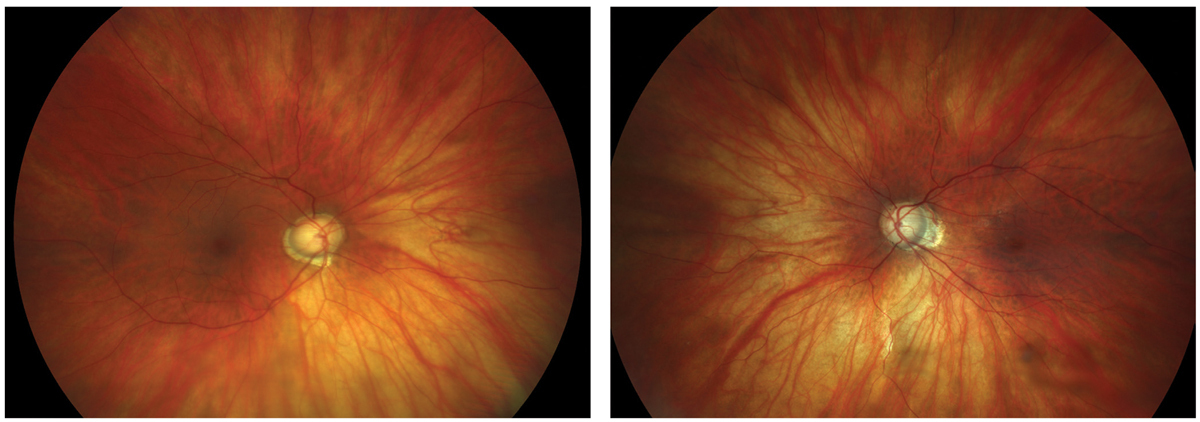
| In this study, contrast sensitivity was associated with chorioretinal structure and vasculature in high myopes. Photo: Sarah B. Klein, OD. Click image to enlarge. |
Researchers recently found that, compared with low to moderate myopic eyes, patients with high myopia have thinner retinal and choroidal thickness, lower retinal vascular density and reduced contrast sensitivity. Moreover, their contrast sensitivity function was correlated with the chorioretinal structure and vasculature.
Eighty-one young subjects were enrolled in this study. They were categorized into the high myopia patient group (n=51) and the low to moderate myopia control group (n=30).
Measures of contrast sensitivity in the high myopia group were significantly reduced compared with those of the control group. The parafoveal and perifoveal retinal thicknesses, deep vascular density and choroidal thickness were also significantly reduced in the high myopia group. Multiple regression analysis revealed that the area under the log of the contrast sensitivity function parameter was significantly correlated with retinal deep vascular density and outer retinal thickness in the parafoveal and perifoveal areas.
“The results suggest that the contrast sensitivity function is a sensitive functional endpoint in simple early-stage high myopia,” the study authors concluded in their paper.
Liu X, Wang Y, Ying X, et al. Contrast sensitivity is associated with chorioretinal thickness and vascular density of eyes in simple early-stage high myopia. Front Med (Lausanne). 2022;9:847817. |