 |
| Genetic testing for keratoconus risk prediction is improving. |
Obtaining very large keratoconus datasets for a genome-wide association study is problematic, as few population studies include Scheimpflug imaging and severe keratoconus is relatively rare. As an alternative to performing genome-wide association studies of keratoconus directly, recent studies of quantitative corneal parameters have proven useful for improving our understanding of the biomechanisms underlying keratoconus. Because patients with keratoconus typically have thinner corneas, corneal central thickness (CCT) is directly relevant. Corneal resistance factor is also significantly decreased in eyes with keratoconus. A recent JAMA Ophthalmology study’s findings suggest an association between a CCT/corneal resistance factor polygenic risk score and improved accuracy in identifying the condition.
This multi-trait genome-wide association study included 105,427 participants from the UK Biobank, 18,307 from the Canadian Longitudinal Study on Aging and 17,803 from European ancestry CCT data from the International Glaucoma Genetics Consortium. A total of 369 corneal resistance factor loci and 233 CCT loci were identified, including 36 novel corneal resistance factor loci and 114 novel CCT loci. The researchers found that 29 corneal resistance factor loci and 24 CCT loci were associated with keratoconus. Polygenic risk scores were constructed using corneal resistance factor- and CCT-associated variants and published keratoconus variants.
“Our findings suggest an association between our polygenic risk score model based on CCT and corneal resistance factor and improved accuracy for identifying keratoconus,” the researchers wrote in their paper. “These findings underscore the power of multi-trait genome-wide association study in identifying new disease-related variants using quantitative traits.”
Brian Chou, OD, of ReVision Optometry, a referral clinic for keratoconus in San Diego, noted that clinicians are wise to recognize that keratoconus manifests within a wide spectrum.
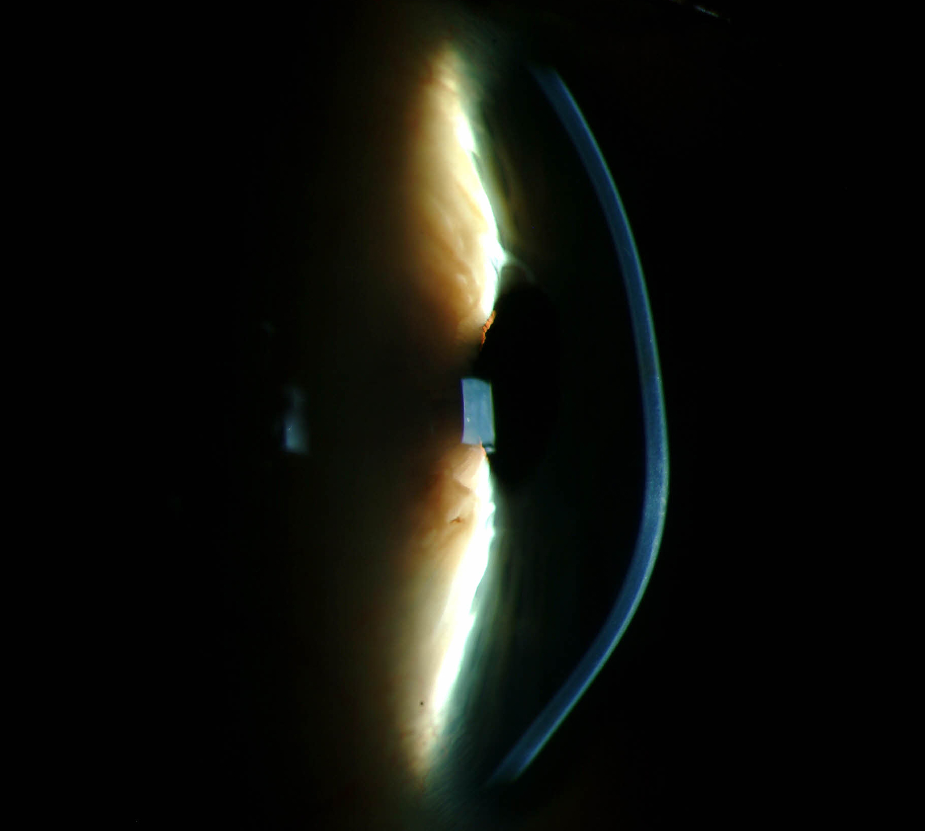
“On one end of severity, keratoconus may be mild enough to escape detection (forme fruste keratoconus), yet on the opposite end, it can be so severe that penetrating keratoplasty is needed,” he noted. “The variability in presentation likely reflects a complex interplay between genetics and environmental influences.”
Dr. Chou says this study “is welcome, since it moves diagnosis of keratoconus from lagging indicators of disease—like Munson's sign, Vogt's striae and apical scarring—toward leading indicators.” Getting ahead in diagnosis enables corneal crosslinking at an earlier stage to slow or halt ectatic progression, he points out. But he also believes there are almost assuredly other important, yet unconfirmed, genetic loci also associated with keratoconus beyond those identified in this study.
“The hope is that genetic testing for keratoconus will advance to the point where it has high clinical validity and utility,” Dr Chou says. “I'm not sure we are there yet, but studies like this can help.”
Ue W, Han X, On JS, et al. Association of novel loci with keratoconus susceptibility in a multitrait genome-wide association study of the UK Biobank database and Canadian Longitudinal Study on Aging. JAMA Ophthalmol. April 21, 2022. [Epub ahead of print]. |