 |
Corneal tomography is integral to guiding treatment decisions in keratoconus, especially for thinner corneas that are sensitive to certain interventions, such as epi-off CXL. Photo: Christine Sindt, OD. Click image to enlarge. |
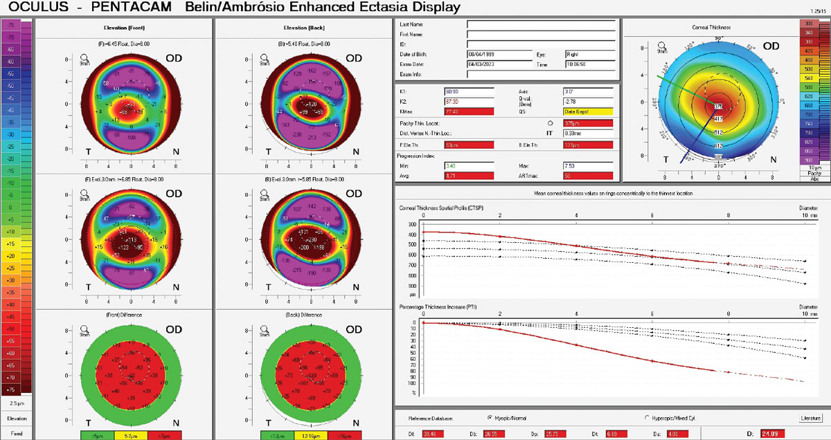
Patients with progressive keratoconus and corneas below 400µm thick are a challenging cohort to manage because these patients may be at risk of UV-mediated endothelial damage in epithelium-off crosslinking (CXL) and increased risk of corneal hydrops. Selecting suitable treatment, therefore, depends on accurate measurement of corneal pachymetric and keratometric parameters and sound repeatability limits. Unfortunately, it’s in just these patients that repeatability of corneal measurements gets increasingly weak.
Researchers recently examined the repeatability of corneal tomographic parameters in patients with keratoconus with advanced corneal thinning in a real-world setting. They found repeatability limits were higher (in other words, worse) in very severe keratoconus (thinnest corneal thickness ≤330µm) vs. severe keratoconus (thickness 331µm to 400µm), both of which were collectively higher than in eyes with moderate thinning (thickness 450µm to 500µm).
Three tomography scans using the Pentacam AXL (Oculus) were obtained from patients with keratoconus with thinnest corneal thickness ≤400µm (sub-400µm group) and compared with those with thickness in the 450µm to 500µm range (450µm+ group). Eyes with previous CXL, intraocular surgery or acute corneal hydrops were excluded. Eyes were age- and gender-matched. The within-subject standard deviations for flat keratometry (K1), steep keratometry (K2), maximal keratometry, astigmatism and thinnest corneal thickness were used to calculate respective repeatability limits.
The sub-400µm group comprised 114 eyes from 114 participants, and the 450µm+ group comprised 114 eyes from 114 participants. In the sub-400µm group, thinnest corneal thickness was amongst the least repeatable parameters (33.92µm) compared with the 450µm+ group (14.32µm). In the sub-400µm group, K1 and K2 of the anterior surface were the most repeatable parameters compared with the 450µm+ group.
“Our study found simulated K1 and K2 of the anterior and posterior corneal surfaces had equivalent or better repeatability than thinnest corneal thickness in both the sub-400µm and 450+µm groups,” the researchers wrote in their paper. “This can be explained by the fact that these parameters represent more global measurements of the corneal surface rather than single-point parameters like thinnest corneal thickness and, thus, are expected to be more repeatable.”
The team suggested that, when assessing patients with advanced thinning (thinnest corneal thickness ≤400µm), clinicians should strive to obtain scans with “OK” quality value if possible and, if unable after several attempts, assess the variability between scans to discern the most likely values of parameters. Using the mean of several measurements—ideally after acquiring a minimum of three scans—may also be helpful. Practitioners should use newer progression analysis systems that incorporate more repeatable parameters.
“This study quantified the repeatability limits of corneal tomographic parameters in keratoconic patients with moderate and advanced corneal thinning, which has implications for the safe application of CXL and identifying disease progression,” they concluded in their paper.
Wadhwa H, Gokul A, Li Y, et al. Repeatability of Scheimpflug based corneal tomography parameters in advanced keratoconus with thin corneas. Eye (Lond). April 19, 2023. [Epub ahead of print]. |