 |
|
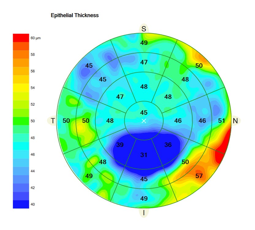
A recent study reported a significant difference between the minimum and maximum epithelial points between stable and keratoconus eyes, with little difference in pachymetry. Photo: Carl Zeiss Meditec. Click image to enlarge. |
Analyzing epithelial changes in eyes with keratoconus (KC) can be helpful in the early diagnosis of the disease, especially when using spectral-domain (SD) OCT imaging. In a recent study, researchers evaluated the corneal epithelium in eyes that showed documented signs of KC progression and determined the correlation of these epithelial parameters with maximum keratometry (Kmax) and pachymetry.
A total of 150 eyes were included: 50 eyes from patients with documented KC progression, 50 eyes with stable KC and 50 controls. The progressive KC group included eyes with an increased Kmax of 1.00D within one year. Epithelial measures of maximum, minimum, superior and inferior values, as well as the difference between minimum and maximum and epithelial standard deviation were considered, obtained from SD-OCT and compared between groups. Measurements of thinnest point and minimum and maximum epithelial points in pachymetry were also recorded.
“The difference between the minimum and maximum epithelial points was significantly different between stable and KC groups, and some cutoffs can potentially differentiate eyes with progression, unlike this difference in pachymetry. This epithelial change seems to be independent of changes in the Kmax and the thinnest pachymetric point of the cornea,” the authors explained. They emphasized that pachymetric map (stromal and epithelial) measurement is not sensitive to structural changes that occur as KC progresses.
They noted that a possible explanation for the role of the more expressive minimum and maximum epithelial difference in eyes with progression is the rapid cell turnover of the corneal epithelium, which is highly reactive to asymmetries in the shape of the underlying stromal surface.
“Direct measurements of the remodeling of the epithelial layer can, therefore, suggest progression,” they added. “This study also confirms that metrics associated with the asymmetric reactive capacity of the epithelium are capable of detecting subtle differences between groups.”
The study authors concluded that “although epithelial thinning measures are helpful in the diagnosis, epithelial variability is the most useful in detecting eyes that are actively progressing compared with stable KC ones. Furthermore, some variability cutoffs can differentiate eyes with progression with relatively high sensitivity. The epithelium minimum and maximum measure can aid monitoring and eventually suggest the indication of corneal crosslinking before significant visual loss.”
Santhiago MR, Stival LR, Araujo DC, et al. Role of corneal epithelial measurements in differentiating eyes with stable keratoconus from eyes that are progressing. Ophthalmology. November 4, 2022. [Epub ahead of print]. |