 |
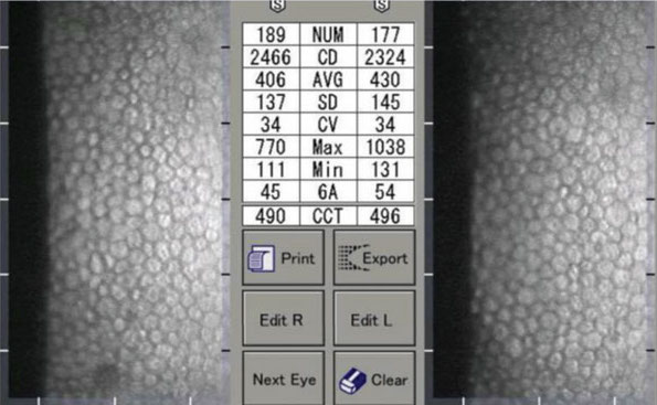
Reduced corneal endothelial density could be expected in eyes that develop myopia. Photo: Daniel Ephstein, OD. Click image to enlarge. |
Little is known about the effect of myopia on the cornea, particularly on the corneal endothelium. To date, few studies have focused on evaluating the correlation between corneal morphology and the anterior chamber. Researchers recently found that greater polymegethism and pleomorphism was observed in healthy eyes with wider mean anterior chamber angle. This implies that the corneal endothelial quality tends to be poorer in eyes with wider anterior chamber angle. These eyes are more likely to be myopic, and myopic corneas may be more fragile and more susceptible to mechanical stress. The team presented their results on the first day of ARVO’s annual meeting in New Orleans.
The study evaluated 272 eyes of 136 Caucasians. Mean age was 46.8, and 61% were men. The endothelial cell density (ECD), coefficient of variation of cell area and hexagonal cell appearance ratio were measured with specular microscopy. Corneal thickness, anterior chamber depth, corneal volume, white-to-white distance and angle width of 12 points were taken by the Pentacam HR (Oculus). Mean anterior chamber angle was the arithmetical mean of the 12 points. The corneal volume was calculated in a 10mm diameter around the corneal apex.
Significant associations were shown between corneal volume, mean anterior chamber angle, white-to-white distance and age with the morphology of the corneal endothelium. Greater polymegathism was found in older individuals, and greater pleomorphism was found in adults with greater white-to-white distance.
Statistical modeling revealed that mean ECD (2673.61 cells/mm2) was positively correlated with corneal volume (59.43mm3). After adjusting for age, it was only negatively correlated with age. The coefficient of variation of cell area (28.6%) was positively correlated with mean anterior chamber angle (35.1º). After adjusting for age, the correlation was stronger. Hexagonal cell appearance ratio (67.5%) was negatively correlated with white-to-white distance (11.8mm) and mean anterior chamber angle, and after adjusting for age, this correlation remained the same.
The researchers noted that the exact mechanism by which myopia could cause corneal endothelial morphology change needs to be further studied.
“Presumably, the endothelial surface area will increase as the axial length elongates and the anterior chamber deepens if the limbal dimension does not change,” they proposed. “Because there is no mitotic activity in the corneal endothelium after birth, it is thus conceivable that the corneal endothelial cells will have to flatten to cover the enlarged surface. Subsequently, a reduced corneal endothelial density could be expected.”
Original abstract content © Association for Research in Vision and Ophthalmology 2023.
Chalkiadaki E, Tsiripidis K, Gartaganis P, et al. Correlations of corneal endothelial morphology with anterior segment parameters in healthy adults. ARVO 2023 annual meeting. |