 |
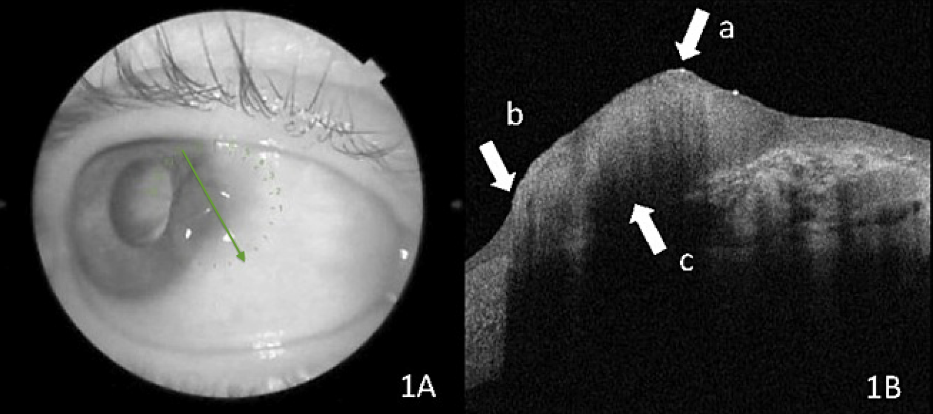
OSSN lesion at the slit lamp and on OCT. The green arrow represents the direction and location of the scans. Arrow A shows a thickened hyperreflective epithelium, arrow B highlights the abrupt transition between abnormal and normal epithelium and arrow C denotes the line of cleavage between the tumor and the surrounding tissue. Photo: Ceyda Başkan/Cureus. Click image to enlarge. |
Accurate diagnosis of ocular surface lesions is imperative to determine the appropriate next step of treatment or removal. Excisional or incisional biopsy is currently the gold standard for reaching a definitive diagnosis; however, this technique is invasive and isn’t always able to detect lesions in the removed tissue.
Several recent studies have examined the potential utility of anterior-segment OCT (AS-OCT) as a noninvasive method for diagnosing and differentiating types of ocular surface squamous neoplasia (OSSN). The technology enables the assessment of morphological and histological features of tissues in vivo. Because these studies have included few cases of pterygium—a benign disease—a group of researchers recently performed their own to further assess the application of AS-OCT in the diagnosis of OSSN, as well as pterygium.
The retrospective study included 21 patients diagnosed with OSSN and 19 with pterygium. AS-OCT measurements were obtained from each patient, and all biopsy materials underwent pathological evaluation.
In patients with OSSN, the researchers found similarities between preoperative AS-OCT images and histopathological specimens. “Both ocular and pathological specimens appeared to have a thicker epithelial layer with a distinct change from healthy to neoplastic epithelium,” they wrote in their paper on the findings.
Patients with pterygium had a normal epithelial thickness and a thickened subepithelial layer based on pathological and AS-OCT images. “The mean epithelial thickness measured with AS-OCT in OSSN patients was found to be 295.3µm, while it was 80.7µm in pterygium patients,” the researchers reported, noting that the difference was statistically significant. “The receiver operating characteristic curve analysis revealed a cut-off value of 97µm for the differential diagnosis of OSSN from pterygium, with a sensitivity of 100% and specificity of 94.7%.”
The findings of this study, as well as those previously conducted, demonstrate that AS-OCT can accurately assist in the detection and differentiation of OSSN and pterygium. “AS-OCT allows clinicians to detect the presence of morphologically suspicious elements in the lesions in a minimally invasive manner,” the researchers explained in their paper. “The number of unnecessary biopsies, surgical excision, cryotherapy and topical chemotherapeutic agent use might be diminished in some patients with the use of AS-OCT.”
Using higher-resolution OCT devices to examine conjunctival lesions in future studies will help develop better documentation of lesion-specific findings to assist clinicians in making surgical decisions for these patients, the researchers concluded.
Başkan C, Kılıcarslan A. How can we diagnose ocular surface squamous neoplasia with optical coherence tomography? Cureus. March 17, 2023. [Epub ahead of print]. |