 |
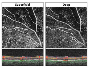
| Superficial and deep capillary plexuses help adjust blood flow in response to metabolic changes. (Photo from different patient) Photo: Carolyn Majcher, OD. Click image to enlarge. |
Previous studies have demonstrated the ability of structures in the retina to regulate blood flow in response to oxygen saturation, as a consistent supply of oxygen is critical for its proper functioning. A group of researchers recently performed a study to better observe the reaction and adaptation mechanism that hyperoxia and hypoxia elicit from superficial and deep capillary plexuses. They found that the capillary plexus plays a large role in the response of retinal circulation to metabolic changes.
The team enrolled 24 subjects in this randomized, double-masked, crossover study to evaluate retinal vascular parameters responding to various levels of oxygen saturation. None of the subjects had clinically significant abnormalities, and only the right eye was observed. On the first of two study days, hyperoxia was induced by breathing 100% oxygen. Within two weeks, a second study day was completed, at which point hypoxia was induced in the same subjects by breathing a mixture of 88% nitrogen and 12% oxygen. Before and during breathing, optical coherence tomography angiography (OCT-A) was used to calculate perfusion density in the superficial vascular plexus (SVP) and the deep capillary plexus (DCP). A dynamic vessel analyzer was used to evaluate the activity of major retinal vessels.
During 100% oxygen breathing, the researchers found study subjects had a significant decrease in DCP perfusion density from 41.7au to 35.6au. However, the same event was not observed in the SVP in response to hyperoxia, which showed no change. On the other hand, during hypoxia, perfusion density in the DCP remained stable while it significantly increased in the SVP from 34.4au. to 37.1au. An increase in retinal vessel diameters was also observed during hypoxia. “Systemic oxygen saturation correlated negatively with perfusion density in the SVP and the DCP and retinal vessel diameters,” the researchers wrote.
“The results of the present study confirm the ability of the retinal microcirculation to adapt its blood flow in response to both hyperoxia and hypoxia with high local resolution,” they concluded based on their findings. “Whereas hyperoxia mainly leads to a constriction in the DCP of the retina, hypoxia leads to vasodilation mainly in the superficial layers. Further, our data show that there is a correlation between systemic oxygen partial pressure and microvascular changes as measured with OCT-A.”
Although these vascular changes can be observed through technology such as OCT-A, the researchers noted, “Little is known about the mechanisms that mediate vasoconstriction and vasodilatation in response to hyperoxia and hypoxia, respectively.” The observations do, however, prove the ability of the retina to self-regulate blood flow in response to metabolic changes.
Hommer M, Kallab M, Sim YC, et al. Effect of hyperoxia and hypoxia on retinal vascular parameters assessed with optical coherence tomography angiography. Acta Ophthalmol. December 8, 2021. [Epub ahead of print]. |