 |
|
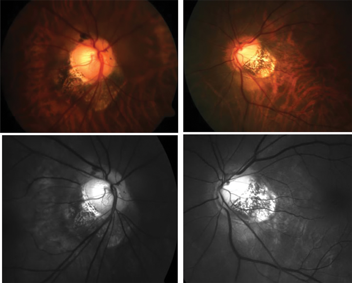
Ocular findings of peripapillary staphyloma in this study cohort included shallower anterior chamber depth, higher IOP, higher myopic spherical equivalent and increased macular deep vessel density. Photo: Andrew Rouse, OD. Click image to enlarge. |
A rare and understudied type of posterior staphyloma found in myopia patients is peripapillary staphyloma, which accounts for between 1.5% and 5% of cases. While studies have confirmed that eyes with posterior staphyloma have worse visual acuity and significant anatomical alterations compared with healthy eyes, little research has been done to determine the specific effects of each condition type.
To help close this gap, researchers recently investigated the vascular and structural characteristics of myopic eyes with peripapillary staphylomas. They identified several findings associated with this type of posterior staphyloma, including worsening of myopia, anatomical alterations of posterior and anterior segments, reductions in the radial peripapillary capillary and macular choroidal perfusion and an increase in macular deep retinal blood flow.
The study examined 82 eyes with myopia, including 41 with peripapillary staphyloma and 41 without, which were selected using propensity score matching based on age and axial length. Compared with controls, eyes with peripapillary staphyloma had a shallower anterior chamber depth (3.6mm vs. 3.8mm), higher intraocular pressure (IOP) (16.6mm Hg vs. 14.5mm Hg) and higher myopic spherical equivalent (-11.5D vs. -9.9D). Corneal curvature and lens thickness did not differ significantly between the two groups.
The researchers also reported in their paper, “Compared with control eyes, increased macular deep vessel density, reduced macular choriocapillaris and radial peripapillary capillary and thinning of the retinal layer, ganglion cell complex, choroidal layer and superior and inferior peripapillary retinal nerve fiber layer were observed in eyes with peripapillary staphyloma.”
Several risk factors of peripapillary staphyloma presence were identified through logistic regression analysis, including higher IOP, rotated optic disc and thinner peripapillary choroid.
“Overall, peripapillary staphyloma may lead to various mechanical changes in the surrounding sclera, retina and choroid, which may explain the different phenotypes,” the researchers concluded in their paper. “Our findings may be helpful in giving new insights into differentiating glaucoma from myopia.”
Nie F, Zhang L, Cao M, et al. Impact of peripapillary staphylomas on the vascular and structural characteristics in myopic eyes: a propensity score matching analysis. Graefes Arch Clin Exp Ophthalmol. January 9, 2023. [Epub ahead of print]. |