 |
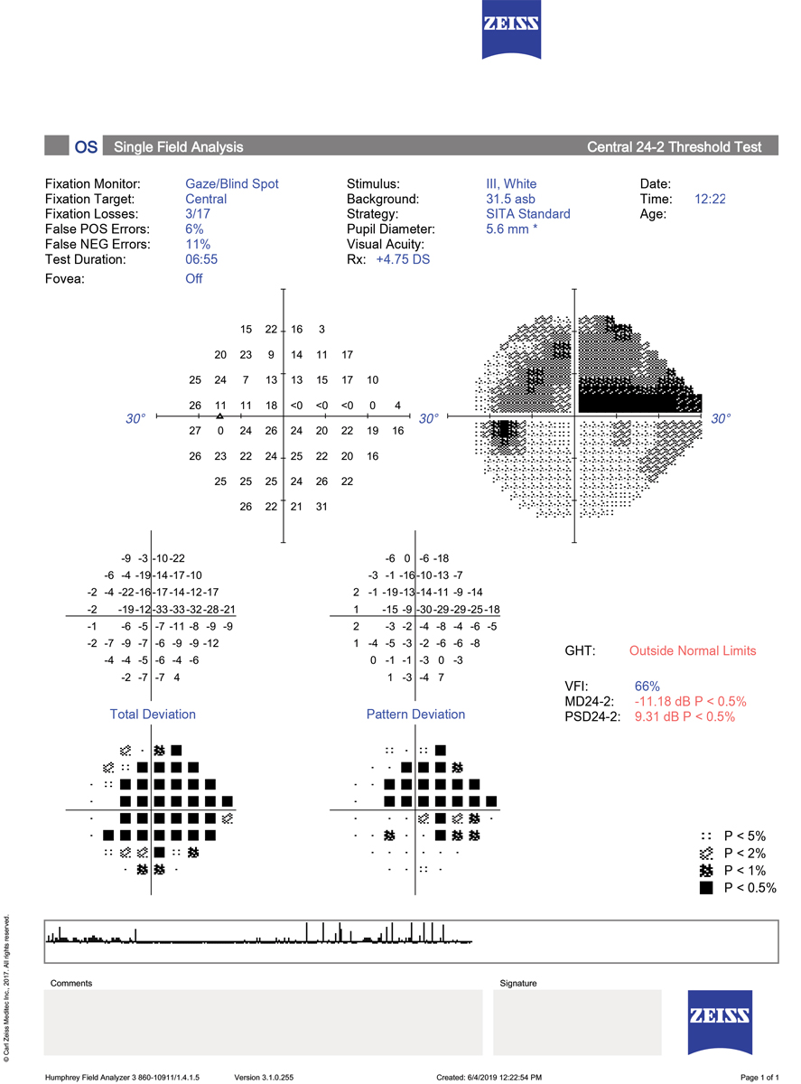
Central visual field defects were observed to increase with glaucoma stage. Photo: Danica Marrelli, OD. Click image to enlarge. |
To better understand glaucoma progression and improve the management of the disease, researchers recently determined the patterns of glaucomatous visual field defects in early, moderate and severe stages of primary open glaucoma using the Glaucoma Staging Application.
They analyzed one visual field of each of the 100 patients included in each group based on the location of the 300 visual defects, involved hemifields and connection to the blind spot.
“The results of this study show that as the severity of the glaucomatous visual field defects increase, their patterns become more central, connected to the physiological blind spot and involving both hemifields,” the authors explained. “We found that even in the early stages of glaucomatous visual field defects, 49% of the defects already occurred in both hemifields, and as the severity increases, the visual field defect deepens and expands, involving the blind spot.”
In the early group, 27% of the visual field defects were connected to the physiological blind spot, 64% in the moderate group and 95% in the severe group.
In the early group, 28% of the defects involved the central 5° of fixation, which prompted the researchers to suggest that the visual field should be carefully monitored in the early stages of glaucoma.
“It is important to note that involvement of both hemifields occurred even in the early stages of functional damage, which may have prognostic values,” the authors explained. “Also, our results show that when the visual field defect is localized in only one hemifield, 62.63% are localized in the superior hemifield.”
Antunes Schiave Germano R, Schiave Germano C, Susanna FN, Junior RS. Patterns of visual field loss in early, moderate and severe stages of open-angle glaucoma. J Glaucoma. January 12, 2022. [Epub ahead of print]. |