 |
|
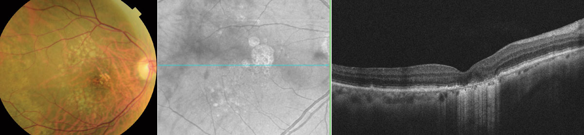
Asian patients with geographic atrophy may have smaller lesion size and slower growth rate than Europeans. Photo: Wendy Harrison, OD, PhD. Click image to enlarge. |
A study recently published in Ophthalmology Retina evaluated differences in geographic atrophy characteristics in Asian (mainly East Asian) and non-Asian individuals. “As potential therapies for geographic atrophy are being investigated mostly in subjects of European descent, understanding of the phenotypes, natural history and risk associated with fast progression are crucial if these therapies are to be considered in Asian populations,” the researchers explained in their study. They reported that geographic atrophy lesions in Asian subjects had smaller baseline size and slower growth rates than those of non-Asian subjects.
The retrospective, multicenter case series included 169 eyes of 144 subjects ≥50 years of age with geographic atrophy secondary to AMD (no neovascularization) and follow-up data spanning at least two years. The researchers characterized geographic atrophy lesions using multimodal imaging (fundus autofluorescence, near infrared and SD-OCT). Roughly half of the study population was Asian (50.9%).
The researchers reported in their paper that Asians had significantly thicker choroids (167µm vs. 134µm) and a significantly lower prevalence of drusen (40.7% vs. 66.3%). They also demonstrated significantly smaller geographic atrophy area at baseline and had fewer foci than non-Asians (3.7mm2 vs. 6.3mm2 on near-infrared imaging, 2.4mm2 vs. 8.4mm2 on fundus autofluorescence, 1.7 vs. 2.7 foci). Geographic atrophy lesion growth rate was slower among Asians than non-Asians (0.7mm2 vs. 1.9mm2 per year on near-infrared imaging, 0.3mm2 vs. 0.2mm2 per year on fundus autofluorescence). Notably, when baseline lesion size was at least 5mm2, ethnic differences were no longer significant.
The researchers concluded that ethnicity, junctional zone fundus autofluorescence pattern, baseline geographic atrophy area and number of geographic atrophy foci were all associated with lesion growth rate. Additionally, they identified subgroups of Asian eyes with fast progression. “Despite the relatively slow growth rate in the overall Asian cohort of 0.7mm2/year, our sensitivity analyses showed faster geographic atrophy growth among Asians with drusen (1.8mm2/year) and those with large baseline GA lesions (2.6mm2/year),” they wrote in their paper. “Asian patients who are fast progressors…may show greater benefit greatly from new geographic atrophy therapies.”
Teo KYC, Fujimoto S, Sadda SR, et al. Geographic atrophy phenotypes in subjects of different ethnicity: Asia-Pacific Ocular Imaging Society Workgroup Report 3. Ophthalmol Retina. 2023. [Epub ahead of print]. |